Sign in
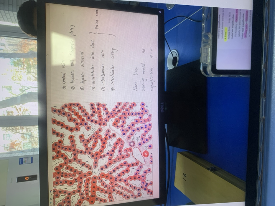
The image displays a histological slide of the liver, stained with Hematoxylin and Eosin (HE) at 10x40 magnification.  Labeled structures include the central vein, hepatic cells (arranged in hepatic plates), hepatic sinusoids, and the portal area, which consists of the interlobular bile duct, vein, and artery. A diagram accompanies the slide, identifying these key features of liver architecture.
Emma🇨🇳💕

Nov 24, 2025, 2:06 AM

Shenyang City, China

Stake attention in this memory

liver histology
microscopic anatomy
hepatic lobule
cell identification
portal triad

The image displays a histological slide of the liver, stained with Hematoxylin and Eosin (HE) at 10x40 magnification. Labeled structures include the central vein, hepatic cells (arranged in hepatic plates), hepatic sinusoids, and the portal area, which consists of the interlobular bile duct, vein, and artery. A diagram accompanies the slide, identifying these key features of liver architecture.

Loading AttnAds…

transactions
revenues
stakers
Earliest
Latest
Highest stake

No transactions found
More from this user

More from Shenyang City

Emma🇨🇳💕

Nov 24, 2025, 2:06 AM

Shenyang City, China

Licensed under Creative Commons Attribution 4.0 International License. You are free to share and adapt this content with proper attribution.