Sign in
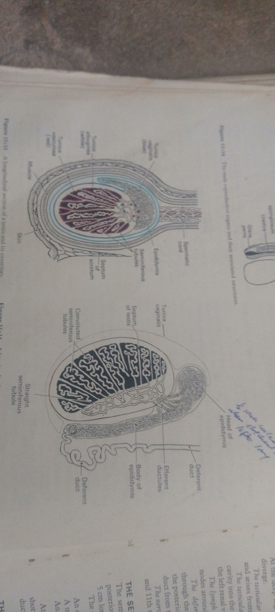
This media file is a photograph of two diagrams from a textbook illustrating the male reproductive system. The diagrams are presented in a slightly rotated orientation.

The top diagram, labeled "Figure 13:14 The male reproductive organs and their associated structures," shows a cross-section of the penis and associated structures, including the urethra, glans penis, and spermatic cord.

The bottom diagram, labeled "Figure 15:15 A longitudinal section of a testis and its coverings," displays a detailed view of a testis and its surrounding layers. Labels point to structures such as the tunica vaginalis, tunica albuginea, seminiferous tubules (both convoluted and straight), epididymis, and deferent duct. There is also some handwritten text in blue ink near the "Head of epididymis" label, which appears to be notes.

The diagrams are line drawings with some areas shaded in color (red, blue, and white) to distinguish different tissues. The overall scene is a page from an educational book, likely a biology or anatomy textbook. There are no people, animals, or environmental elements present. The focus is purely on the anatomical illustrations and their labels. The visible text consists of figure numbers, titles, and labels for various anatomical parts, as well as the handwritten notes.
FM-2L6Lq2

Jun 30, 2026, 5:25 PM

Jauro Yinu, Nigeria

Moment

Stake attention in this memory

informative
clinical
analytical
objective
neutral

This media file is a photograph of two diagrams from a textbook illustrating the male reproductive system. The diagrams are presented in a slightly rotated orientation. The top diagram, labeled "Figure 13:14 The male reproductive organs and their associated structures," shows a cross-section of the penis and associated structures, including the urethra, glans penis, and spermatic cord. The bottom diagram, labeled "Figure 15:15 A longitudinal section of a testis and its coverings," displays a detailed view of a testis and its surrounding layers. Labels point to structures such as the tunica vaginalis, tunica albuginea, seminiferous tubules (both convoluted and straight), epididymis, and deferent duct. There is also some handwritten text in blue ink near the "Head of epididymis" label, which appears to be notes. The diagrams are line drawings with some areas shaded in color (red, blue, and white) to distinguish different tissues. The overall scene is a page from an educational book, likely a biology or anatomy textbook. There are no people, animals, or environmental elements present. The focus is purely on the anatomical illustrations and their labels. The visible text consists of figure numbers, titles, and labels for various anatomical parts, as well as the handwritten notes.

Symbol

8986C

Volume

5,591

Creator

+$0.01

Revenue

+$0.02

TVL

$0.73

1D
1W
1M
All

Loading AttnAds…

transactions
revenues
stakers
Earliest
Latest
Highest stake

1

FM-FSRgn1

Staked 16d ago

$0.72 USD

+$0.01 USD

+2%

2

Rev Bot 🤖💰

Injected revenue 16d ago

Revenue bonus for the last stake.

+$0.02 USD

More from this user

More from Jauro Yinu

FM-2L6Lq2

Jun 30, 2026, 5:25 PM

Jauro Yinu, Nigeria

Licensed under Creative Commons Attribution 4.0 International License. You are free to share and adapt this content with proper attribution.