Stake attention in this memory
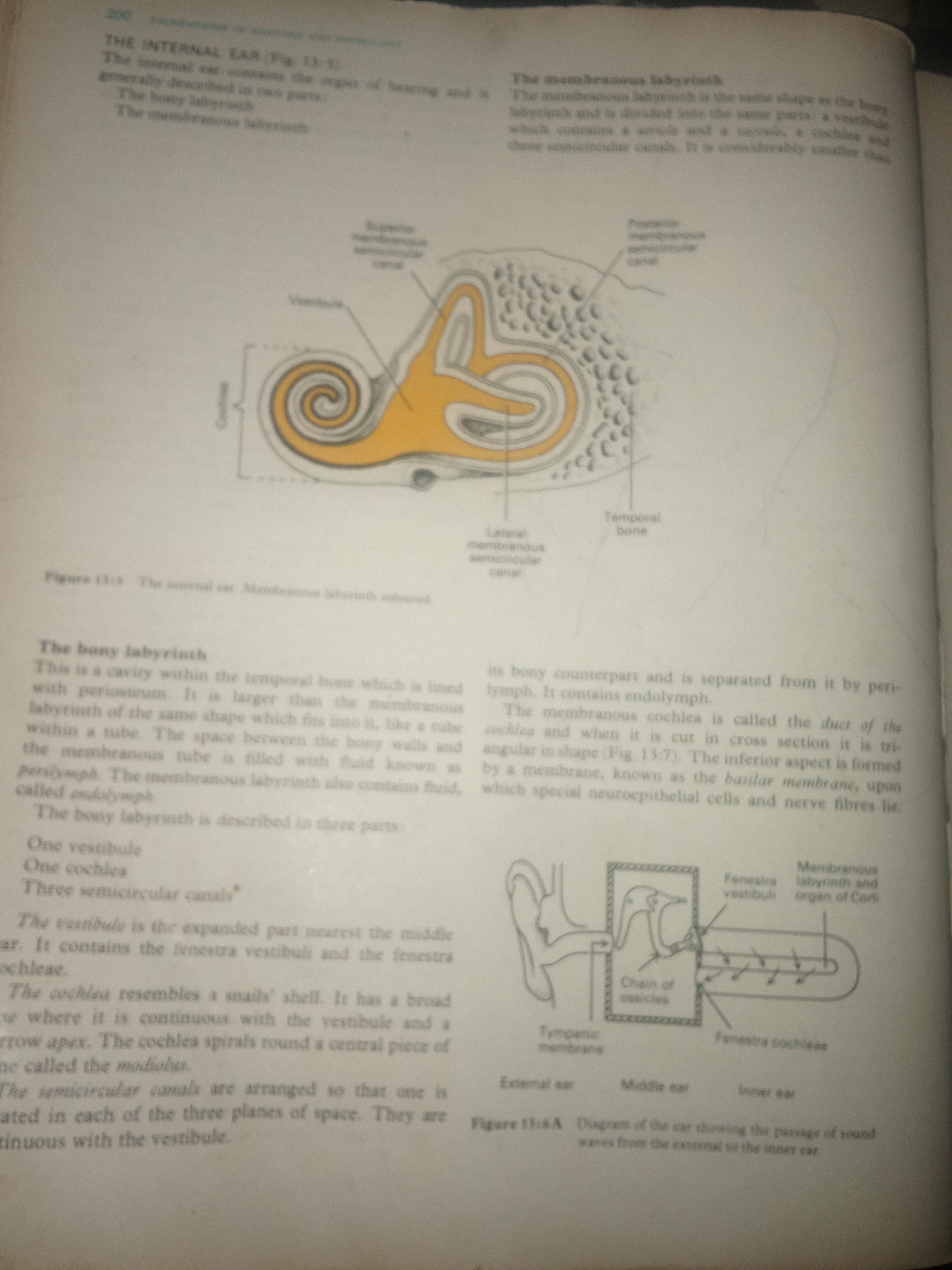
The image shows a two-page spread from a biology textbook. The left page discusses "The bony labyrinth" and "The membranous labyrinth" within the context of the internal ear. There is a diagram illustrating the internal ear, with labels such as "Superior semicircular canal," "Vestibule," "Cochlea," and "Temporal bone." The right page continues the discussion of the membranous labyrinth, explaining its structure and relation to the bony labyrinth. It also describes the cochlea and semicircular canals. Below this text, there is a diagram labeled "Figure 13:6A Diagram of the ear showing the passage of sound waves from the external to the inner ear," which depicts the external ear, middle ear, and inner ear. The text is predominantly scientific and descriptive, providing anatomical and functional information about the human ear. There are no people visible in the image. The setting is a book, likely a classroom or study environment, given the educational nature of the content. The lighting appears to be standard indoor lighting, and there are no indicators of time of day or weather. The focus is on biological illustration and textual explanation. Visible text includes chapter titles, section headings, descriptive text, labels for anatomical parts in the diagrams, and figure captions. The text is in English.
Loading AttnAds…
No transactions found