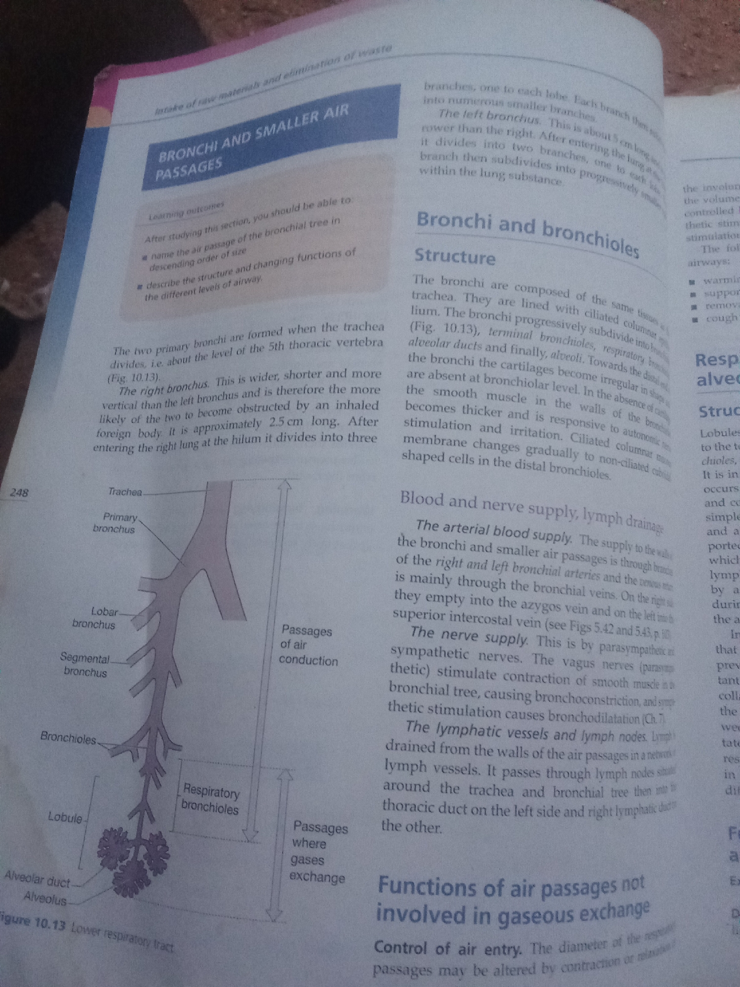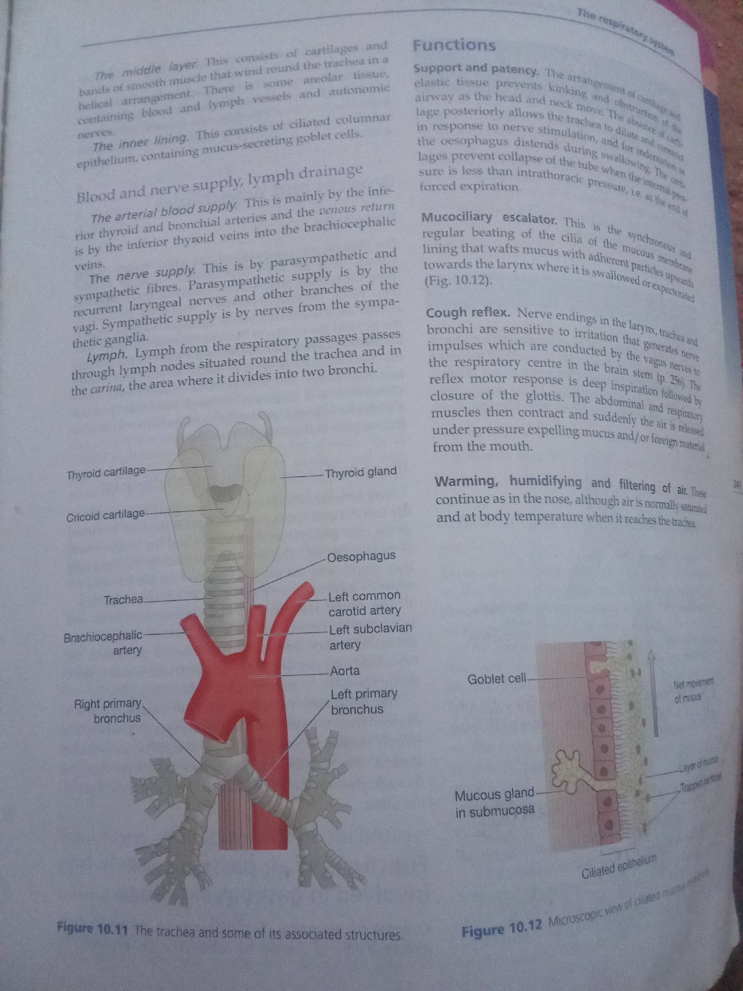
Stake attention in this memory
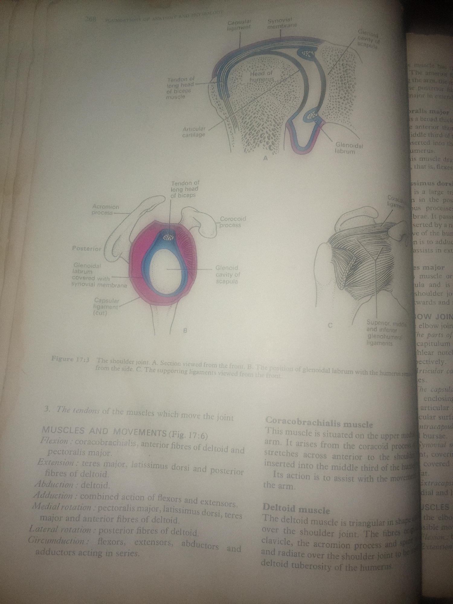
The image is a page from an anatomy textbook illustrating the shoulder joint. It features several diagrams and accompanying text. The main subjects are anatomical illustrations of the shoulder joint, labeled with various parts such as the "head of humerus," "glenoid cavity of scapula," "glenoidal labrum," and "tendon of long head of biceps muscle." There are three diagrams labeled A, B, and C, showing different views and aspects of the joint, including a cross-section, a frontal view, and a posterior view showing ligaments. The text describes the anatomy and movements of the shoulder joint, detailing muscles involved in flexion, extension, abduction, adduction, medial rotation, lateral rotation, and circumduction. Specific muscles like the "coracobrachialis muscle" and "deltoid muscle" are also described, including their origins, insertions, and actions. Notable details include the use of anatomical terminology and the clear, labeled diagrams that are typical of educational material. The "Figure 17:3" designation suggests this is part of a larger work. The image appears to be printed on paper, with a slightly aged look. The text is in English. There are no people or animals depicted. The setting is a printed page from a textbook, with no discernible time of day, weather, or location cues beyond the subject matter of human anatomy. Emotions are not applicable as there are no living beings depicted.
No transactions found