
Sign in
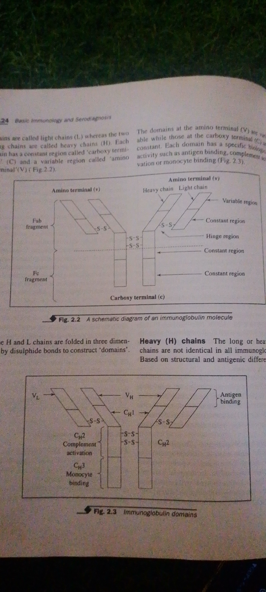
The image displays two diagrams illustrating the structure of immunoglobulin molecules, commonly known as antibodies. These diagrams are from a textbook, as indicated by the chapter number "24 Basic Immunology and Serodiagnosis" and figure captions "Fig. 2.2 A schematic diagram of an immunoglobulin molecule" and "Fig. 2.3 Immunoglobulin domains."

The first diagram (Fig. 2.2) shows a schematic representation of an immunoglobulin molecule, depicting both a light chain and a heavy chain. It labels the "Amino terminal (v)" and "Carboxy terminal (c)" regions, as well as the "Fab fragment" and "Fc fragment." Disulfide bonds ("S-S") are indicated, showing how the chains are linked.

The second diagram (Fig. 2.3) provides a more detailed view of immunoglobulin domains. It labels the light chain variable region (VL) and heavy chain variable region (VH), and the constant regions of the heavy chain (CH1, CH2, CH3). It also highlights the "Antigen binding" site and indicates functions for CH2 ("Complement activation") and CH3 ("Monocyte binding"). The terms "Heavy chain" and "Light chain" are also present, along with labels for "Variable region," "Constant region," and "Hinge region."

The text surrounding the diagrams discusses the structure and function of these chains and their domains, including the role of disulfide bonds in folding and the biological activities associated with different domains. The diagrams are line drawings and do not contain any elements suggesting a specific time of day, weather, or geographical location. The context is purely academic, related to the study of immunology.
FM-2L6Lq2

Jun 30, 2026, 3:40 PM

Jalingo, Nigeria

Moment

Stake attention in this memory

informative
educational
scientific
diagrammatic
analytical

The image displays two diagrams illustrating the structure of immunoglobulin molecules, commonly known as antibodies. These diagrams are from a textbook, as indicated by the chapter number "24 Basic Immunology and Serodiagnosis" and figure captions "Fig. 2.2 A schematic diagram of an immunoglobulin molecule" and "Fig. 2.3 Immunoglobulin domains." The first diagram (Fig. 2.2) shows a schematic representation of an immunoglobulin molecule, depicting both a light chain and a heavy chain. It labels the "Amino terminal (v)" and "Carboxy terminal (c)" regions, as well as the "Fab fragment" and "Fc fragment." Disulfide bonds ("S-S") are indicated, showing how the chains are linked. The second diagram (Fig. 2.3) provides a more detailed view of immunoglobulin domains. It labels the light chain variable region (VL) and heavy chain variable region (VH), and the constant regions of the heavy chain (CH1, CH2, CH3). It also highlights the "Antigen binding" site and indicates functions for CH2 ("Complement activation") and CH3 ("Monocyte binding"). The terms "Heavy chain" and "Light chain" are also present, along with labels for "Variable region," "Constant region," and "Hinge region." The text surrounding the diagrams discusses the structure and function of these chains and their domains, including the role of disulfide bonds in folding and the biological activities associated with different domains. The diagrams are line drawings and do not contain any elements suggesting a specific time of day, weather, or geographical location. The context is purely academic, related to the study of immunology.

Symbol

8CBD1

Volume

7,350

Creator

+$0.01

Revenue

+$0.02

TVL

$1.11

1D
1W
1M
All

Loading AttnAds…

transactions
revenues
stakers
Earliest
Latest
Highest stake

1

FM-FSRgn1

Staked 1mo ago

$1.11 USD

+$0.00 USD

+0.4%

2

Rev Bot 🤖💰

Injected revenue 1mo ago

Revenue bonus for the last stake.

+$0.02 USD

More from this user

More from Jalingo

FM-2L6Lq2

Jun 30, 2026, 3:40 PM

Jalingo, Nigeria

Licensed under Creative Commons Attribution 4.0 International License. You are free to share and adapt this content with proper attribution.