Stake attention in this memory
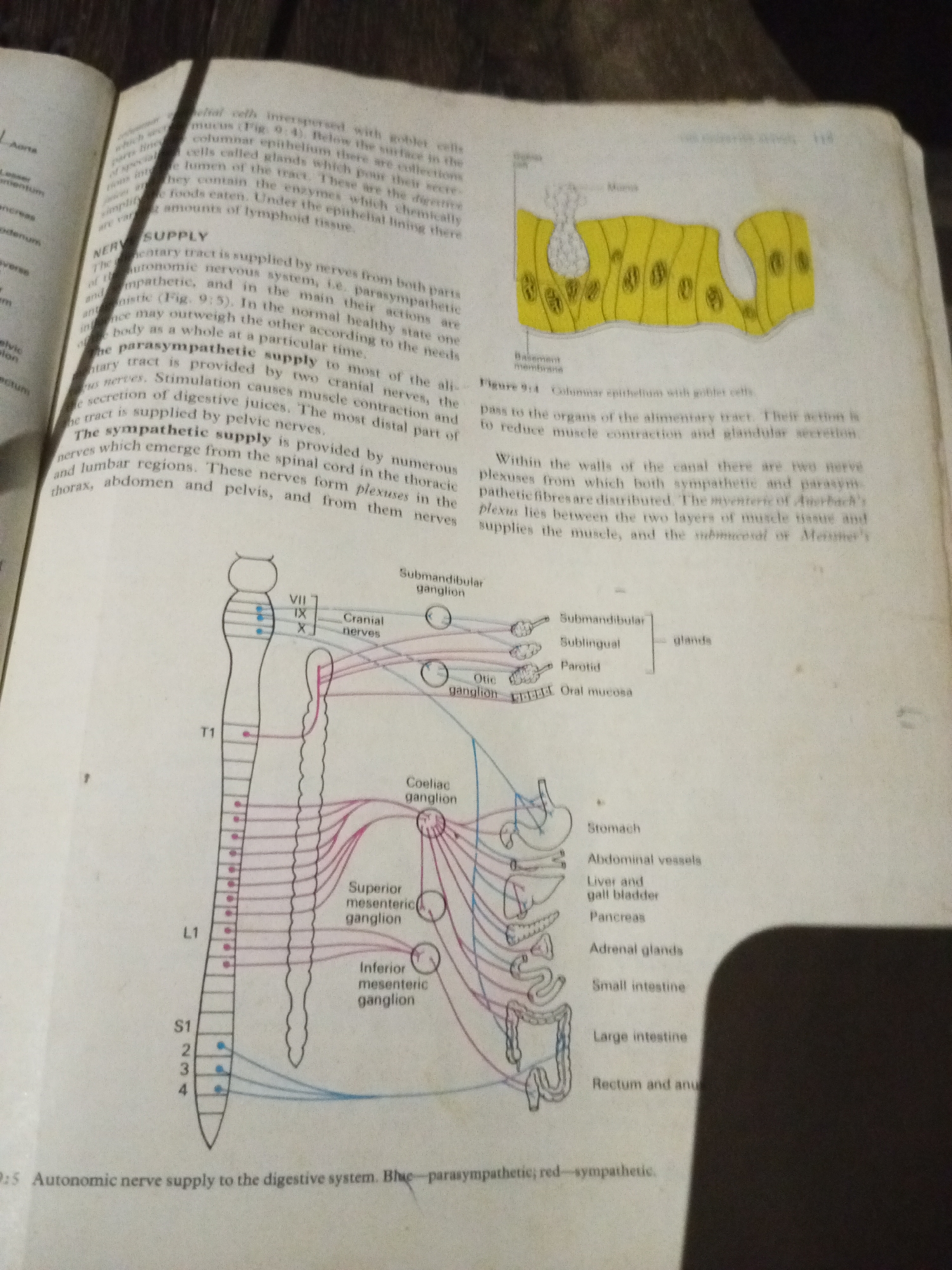
The image contains a page from a textbook. On the left side, there is a diagram illustrating the autonomic nerve supply to the digestive system. It shows the spinal cord with labels indicating thoracic (T1) and lumbar (L1, S1-4) segments, connected by red and blue lines to various ganglia (Superior mesenteric, Inferior mesenteric, Coeliac) and then to organs like the stomach, abdominal vessels, liver and gall bladder, pancreas, adrenal glands, small intestine, large intestine, and rectum and anus. The blue lines represent parasympathetic supply and the red lines represent sympathetic supply. To the right of the diagram is an illustration of columnar epithelium with goblet cells, labeled as Figure 9.4. Below this figure, text describes the functions of the epithelium and the nerve supply to the alimentary tract, mentioning parasympathetic and sympathetic supply. The text details how these nerves originate from the cranial nerves and spinal cord, forming plexuses within the digestive tract walls. The page also includes some text from the surrounding pages of the textbook, partially visible on the left and top edges. The overall environment appears to be a flat surface, likely a table, with shadows cast across the page, indicating overhead lighting. The image is taken from a slightly overhead angle. There are no people or identifiable locations other than those depicted within the textbook content.
Symbol
8E1B2
Volume
11,550
Creator
+$0.17
Revenue
+$0.24
TVL
$14.71
2
Rev Bot 🤖💰
Injected revenue 53m ago
“Revenue bonus for the last stake.”
+$0.26 USD