
Stake attention in this memory
medical imaging
bone necrosis
X-ray
CT scan
MRI
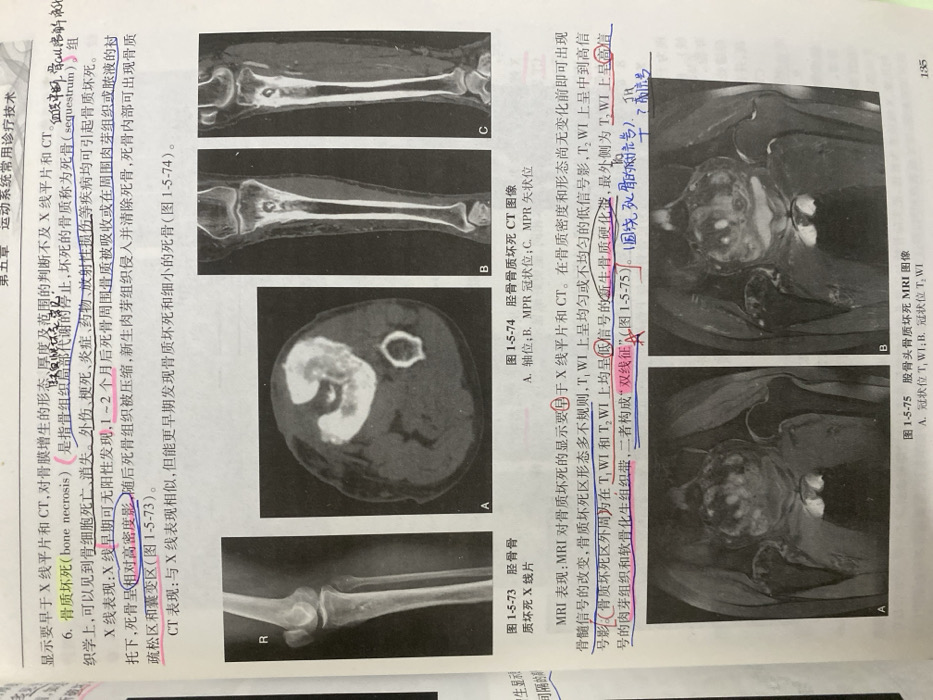
The image presents diagnostic techniques for bone necrosis, including X-ray, CT, and MRI. X-ray images may show increased density in early stages, followed by areas of bone loss. CT scans are similar to X-rays but can detect earlier and smaller necrotic regions. MRI is the most sensitive, showing changes in bone marrow signal before density changes are visible. A "double line" sign is noted, consisting of granulation tissue and a sclerotic rim on T2-weighted images. The images show examples of bone necrosis in the tibia and femoral head.
Loading AttnAds…
transactions
revenues
stakers
Earliest
Latest
Highest stake
No transactions found