Stake attention in this memory
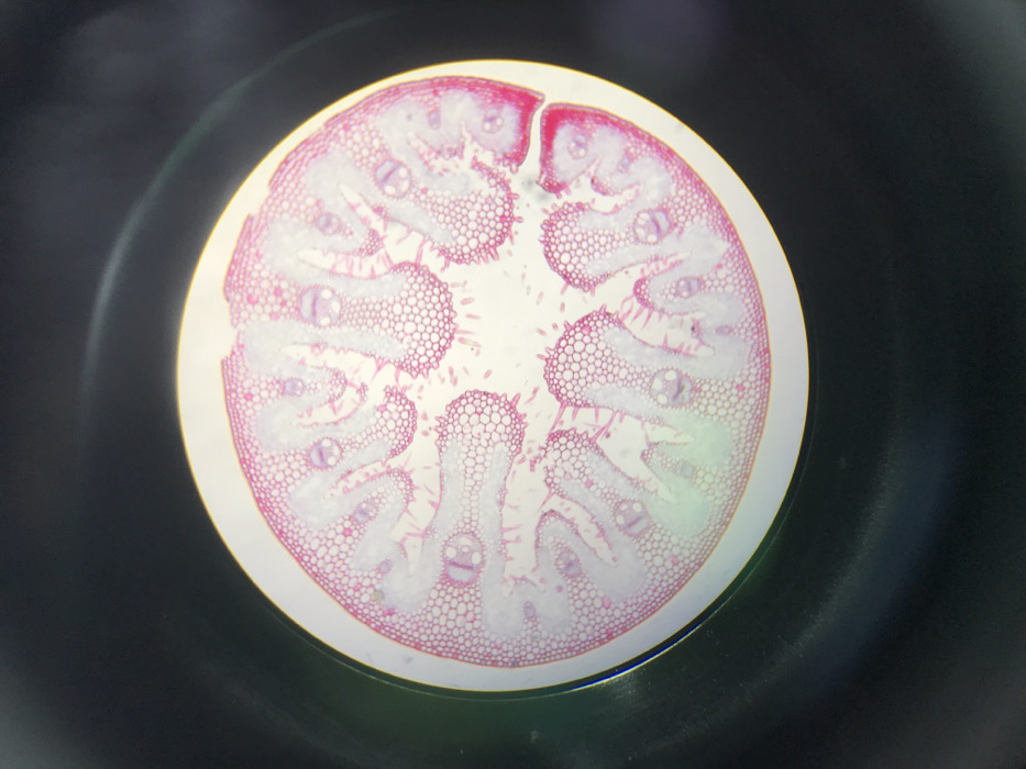
The image displays a circular microscopic view of a stained biological cross-section, occupying approximately 90% of the frame against a dark, out-of-focus background. The specimen, likely plant tissue, is predominantly stained in various shades of pink and white. An outer epidermal layer, characterized by tightly packed, small cells, exhibits a distinct magenta coloration. Beneath this, the tissue forms an undulating, wave-like structure with numerous vascular bundles embedded within a lighter pink to white ground tissue. These bundles often project inwards or outwards, some appearing to have more intensely stained caps. Fine cellular detail is visible, with varying cell sizes and arrangements throughout the section. No individuals or active processes are depicted. This microscopic observation is documented from Midrand, South Africa.
No transactions found