Sign in
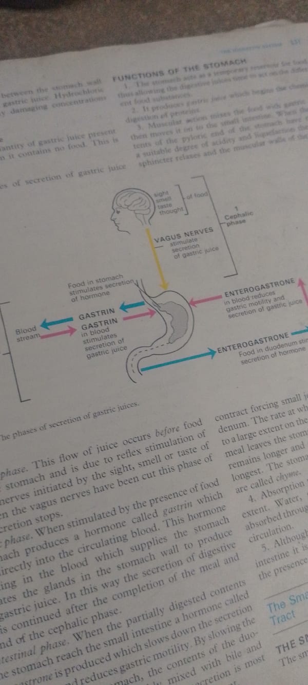
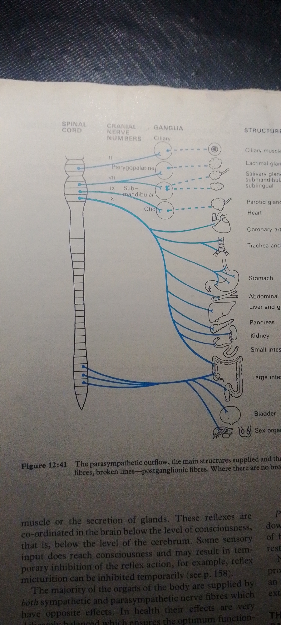
This media file is a diagram illustrating the parasympathetic outflow of the nervous system, detailing the structures supplied by these nerve fibers. The diagram originates from a book, as indicated by the presence of text and figure captions.

The central focus of the image is a schematic representation of the spinal cord on the left, with nerve pathways branching out to various organs on the right. These organs are depicted as line drawings and include the eye (ciliary muscle, lacrimal gland), salivary glands, parotid gland, heart, coronary artery, trachea, stomach, liver, pancreas, kidney, intestines (small and large), bladder, and sex organs.

The diagram visually connects cranial nerve numbers (III, VII, IX, X) and associated ganglia (Ciliary, Pterygopalatine, Submandibular, Otic) to these organs, differentiating between preganglionic (solid lines) and postganglionic (broken lines) fibers.

Visible text includes labels for the parts of the nervous system and the organs, as well as a figure caption "Figure 12:41 The parasympathetic outflow, the main structures supplied and the fibres, broken lines—postganglionic fibres. Where there are no bro". There are also snippets of descriptive text from the accompanying book page, discussing reflexes and the coordination of the body's organs.

The image is a static, illustrative diagram, not depicting any people, a specific scene, or an event in a real-world location. The setting is the page of a book, likely in a medical or biology textbook. There are no cues regarding time of day, weather, or emotions.
FM-2L6Lq2

Jun 30, 2026, 4:54 PM

Jauro Yinu, Nigeria

Moment

Stake attention in this memory

curious
educational
informative
analytical
detailed

This media file is a diagram illustrating the parasympathetic outflow of the nervous system, detailing the structures supplied by these nerve fibers. The diagram originates from a book, as indicated by the presence of text and figure captions. The central focus of the image is a schematic representation of the spinal cord on the left, with nerve pathways branching out to various organs on the right. These organs are depicted as line drawings and include the eye (ciliary muscle, lacrimal gland), salivary glands, parotid gland, heart, coronary artery, trachea, stomach, liver, pancreas, kidney, intestines (small and large), bladder, and sex organs. The diagram visually connects cranial nerve numbers (III, VII, IX, X) and associated ganglia (Ciliary, Pterygopalatine, Submandibular, Otic) to these organs, differentiating between preganglionic (solid lines) and postganglionic (broken lines) fibers. Visible text includes labels for the parts of the nervous system and the organs, as well as a figure caption "Figure 12:41 The parasympathetic outflow, the main structures supplied and the fibres, broken lines—postganglionic fibres. Where there are no bro". There are also snippets of descriptive text from the accompanying book page, discussing reflexes and the coordination of the body's organs. The image is a static, illustrative diagram, not depicting any people, a specific scene, or an event in a real-world location. The setting is the page of a book, likely in a medical or biology textbook. There are no cues regarding time of day, weather, or emotions.

Symbol

93683

Volume

5,857

Creator

+$0.01

Revenue

+$0.01

TVL

$0.78

1D
1W
1M
All

Loading AttnAds…

transactions
revenues
stakers
Earliest
Latest
Highest stake

1

FM-FSRgn1

Staked 16d ago

$0.78 USD

+$0.01 USD

+0.7%

2

Rev Bot 🤖💰

Injected revenue 16d ago

Revenue bonus for the last stake.

+$0.01 USD

More from this user

More from Jauro Yinu

FM-2L6Lq2

Jun 30, 2026, 4:54 PM

Jauro Yinu, Nigeria

Licensed under Creative Commons Attribution 4.0 International License. You are free to share and adapt this content with proper attribution.