
Stake attention in this memory
microscopic image
histology
liver tissue
cells
staining
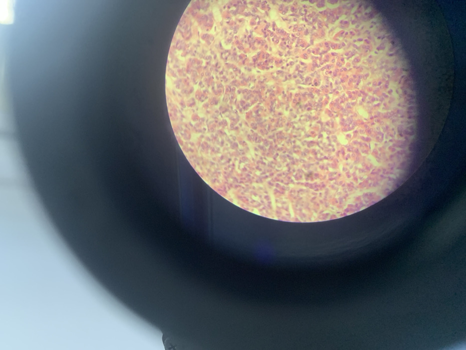
A microscopic view displays a tissue sample, likely stained with eosin, presenting a field of pink-hued cells. The cellular structure appears dense and somewhat disorganized, with visible nuclei and cytoplasm. Connective tissue or interstitial space is present, appearing lighter in color. The image is captured through a microscope lens, with a circular field of view partially obscured by the instrument's components. Overall, it exhibits characteristics of cellular detail in histological analysis.
Loading AttnAds…
transactions
revenues
stakers
Earliest
Latest
Highest stake
No transactions found