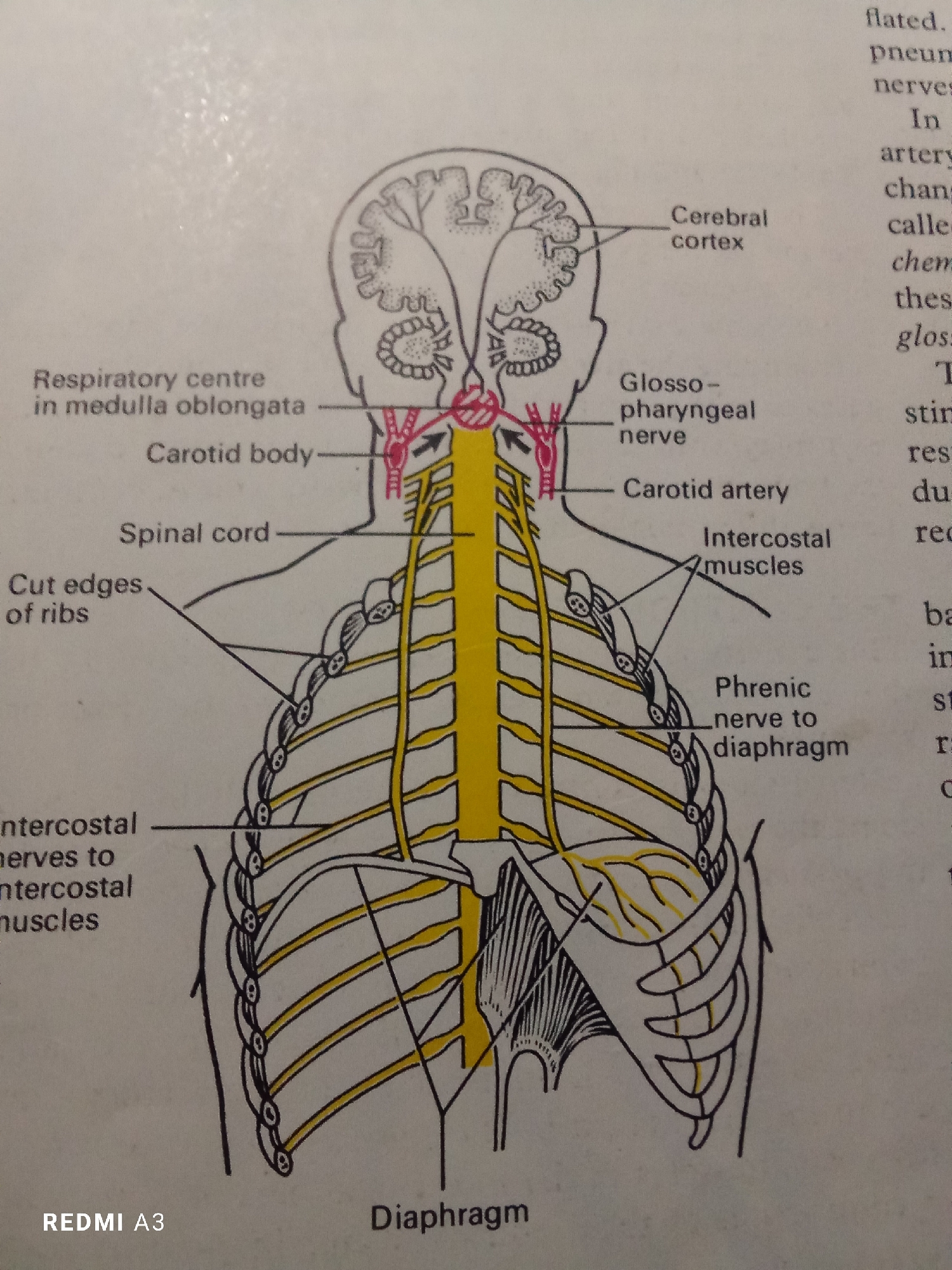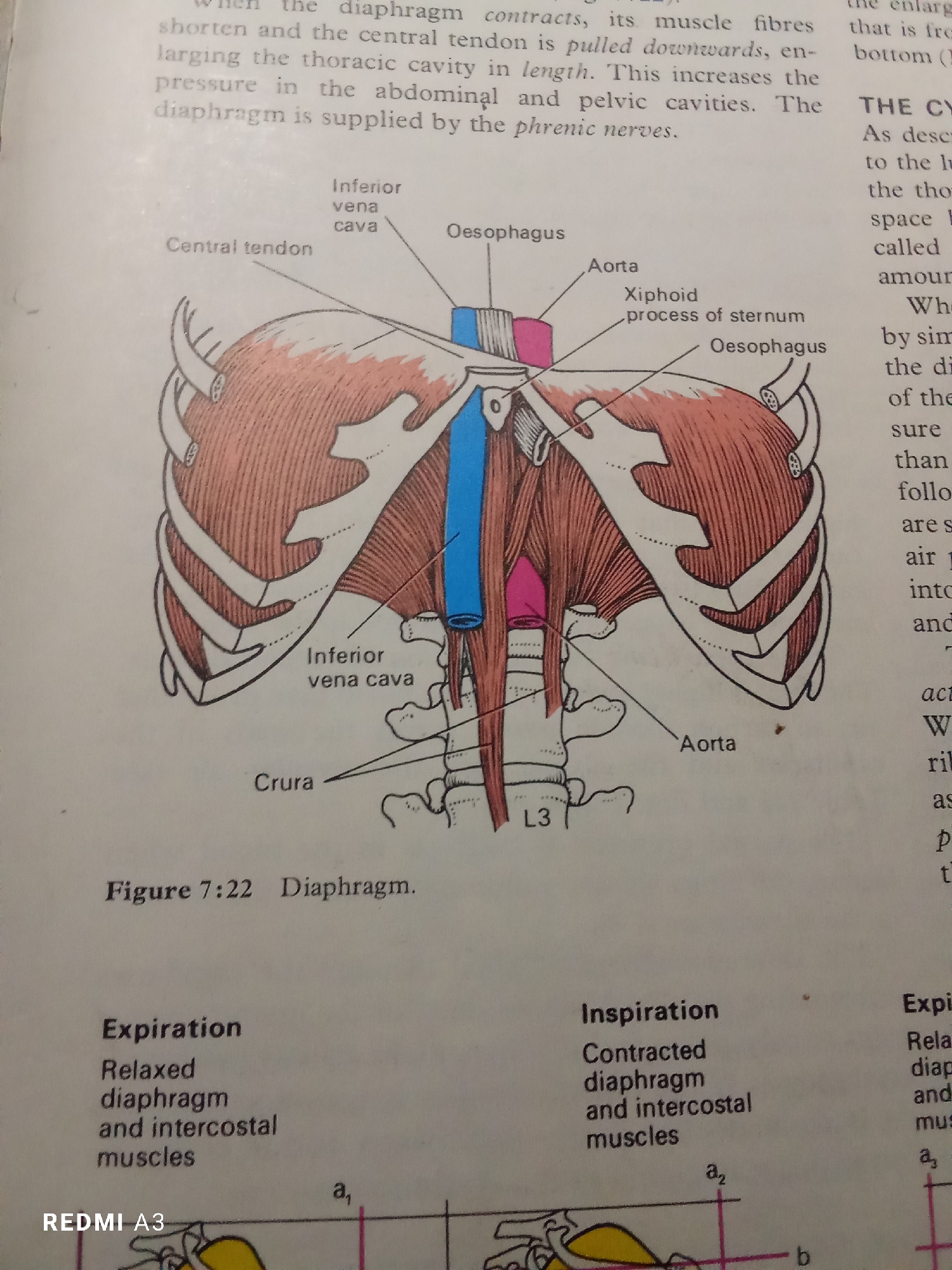
Stake attention in this memory
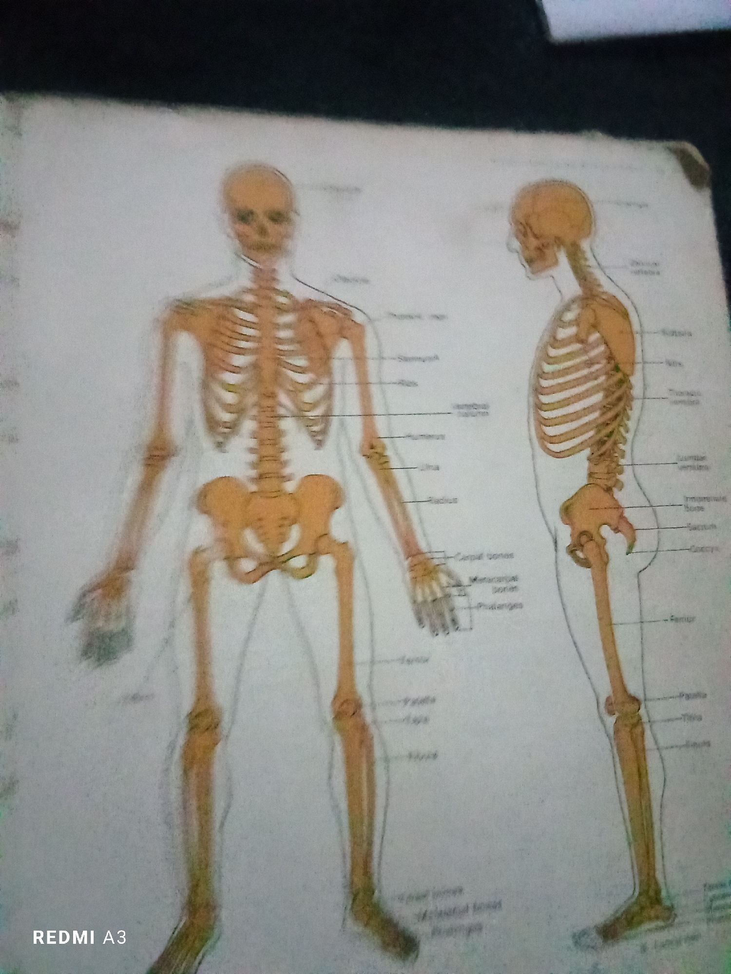
This media file displays an illustration of the human skeleton, presented in both anterior and lateral views. The skeleton is colored in shades of orange and yellow, with labels pointing to various bones. **Subjects:** The primary subjects are two diagrams of the human skeletal system. No people are depicted, only the bony structures. **Activity or event:** The image is a static illustration, likely from an educational textbook or chart, intended to depict anatomical information. There is no activity or event taking place. **Notable details:** * The anterior view shows the skeleton facing forward, with the rib cage, vertebral column, pelvis, arms, and legs clearly visible. * The lateral view shows the skeleton from the side, highlighting the curvature of the spine and the structure of the skull and rib cage. * Numerous labels are present, identifying specific bones such as "Skull," "Vertebrae," "Ribs," "Humerus," "Ulna," "Radius," "Carpal bones," "Metacarpal bones," "Phalanges," "Femur," "Patella," "Tibia," and "Fibula." * The illustration is set against a white background. * The image quality is somewhat blurry. **Visible text:** * "REDMI A3" is visible in the bottom left corner of the image. This is likely a watermark from the device used to capture the photo. * Various anatomical labels are present throughout the illustration, as mentioned above.
No transactions found