
Stake attention in this memory
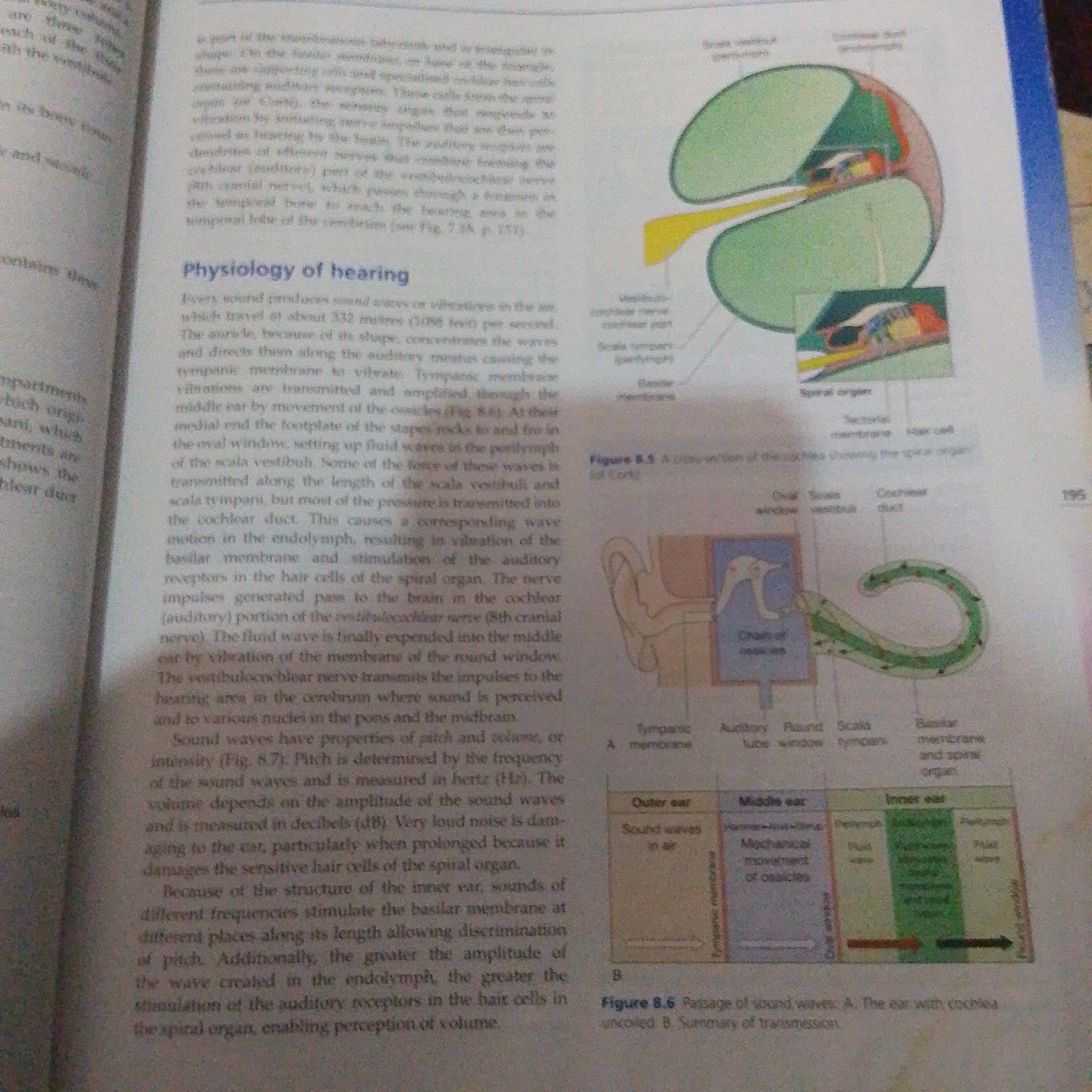
The media file is a close-up photograph of an open textbook page. The primary subject is the printed text and accompanying diagrams illustrating the physiology of hearing. The text discusses how sound waves travel through the ear, detailing the roles of different parts like the tympanic membrane, ossicles, and cochlea. Two diagrams are visible. The larger diagram, labeled "Figure 8.5 A cross-section of the cochlea showing the spiral organ," depicts a cross-section of the cochlea with various parts labeled, including the "Scala tympani," "Scala vestibuli," "tectorial membrane," "hair cell," and "spiral organ." A smaller inset diagram shows a magnified view of a section of the cochlea. Below this, "Figure 8.6 Passage of sound waves" presents a simplified diagram of the ear divided into "Outer ear," "Middle ear," and "Inner ear," summarizing the transmission of sound. The text describes the process of hearing, including the vibration of membranes, the role of fluid waves in the inner ear, and the stimulation of auditory receptors. It also touches upon the properties of sound waves, such as pitch and volume, and how very loud noise can damage the ear. There are no people, animals, or discernible weather or time of day cues. The setting is clearly an educational context, likely a classroom or library, where a textbook is being studied. The visible text is entirely in English and relates to scientific or medical information about auditory function. There is also a page number "195" visible on the right side of the page.
Loading AttnAds…
No transactions found