
Stake attention in this memory
anatomy
bone
humerus
muscles
diagram
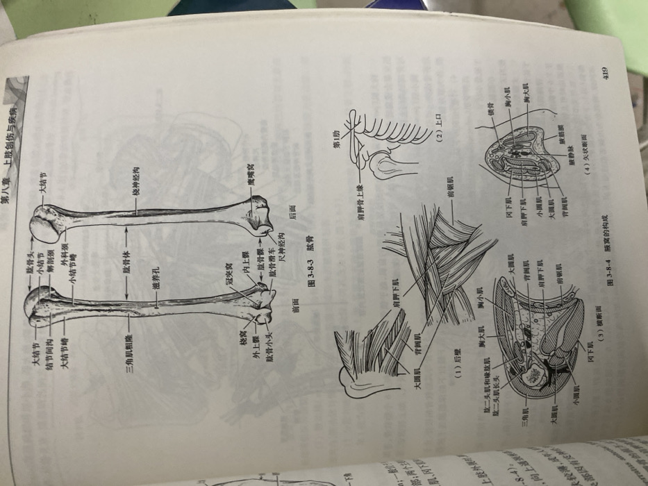
The image displays anatomical drawings from a medical textbook. One section illustrates the humerus bone from anterior and posterior views, labeling structures like the greater tubercle, lesser tubercle, anatomical neck, surgical neck, deltoid tuberosity, radial groove, and condyles. Additional illustrations depict the axilla's composition, showing muscles (teres major, latissimus dorsi, serratus anterior, subscapularis), veins, and fascia. The layout includes sagittal and transverse sections of the axilla, offering a comprehensive anatomical overview. The diagram also shows a view of the first rib and scapula.
Loading AttnAds…
transactions
revenues
stakers
Earliest
Latest
Highest stake
No transactions found