Sign in
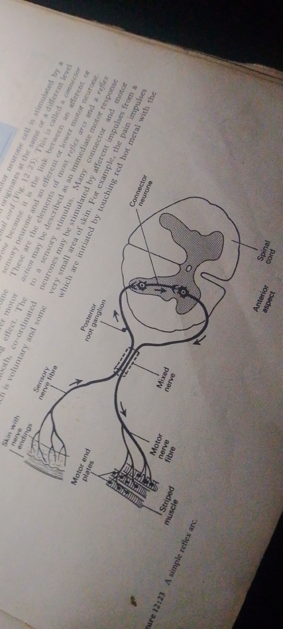
The image shows a diagram from a textbook illustrating "A simple reflex arc." The diagram depicts the neural pathway involved in a reflex action, starting from sensory nerve endings in the skin, traveling through a sensory neuron, a connector neuron (also called an interneuron) within the spinal cord, and finally to a motor neuron that innervates a striped muscle.

The diagram includes labels such as "Skin with nerve endings," "Sensory nerve fibre," "Posterior root ganglion," "Connector neurone," "Spinal cord," "Anterior aspect," "Mixed nerve," "Motor nerve fibre," and "Motor end plates." The spinal cord is shown in cross-section, with the gray matter centrally located in a butterfly or H-shape, and the white matter surrounding it. Arrows indicate the direction of nerve impulse transmission.

Accompanying the diagram is text that explains the function of connector neurons and reflex arcs. It mentions that reflex arcs are the elements of most reflex actions and can be initiated by sensory stimuli, providing an example of pain impulses from touching a hot object. The text also defines a connector neuron as a link between an afferent (sensory) and an efferent (motor) neuron. The image is a scan from a book, indicated by the text and the page edges visible. There are no people or specific environmental cues like time of day or weather. The overall setting is an educational context, likely a biology or physiology textbook.
FM-2L6Lq2

Jun 30, 2026, 4:52 PM

Jauro Yinu, Nigeria

Moment

Stake attention in this memory

analytical
studious
focused
intellectual
curious

The image shows a diagram from a textbook illustrating "A simple reflex arc." The diagram depicts the neural pathway involved in a reflex action, starting from sensory nerve endings in the skin, traveling through a sensory neuron, a connector neuron (also called an interneuron) within the spinal cord, and finally to a motor neuron that innervates a striped muscle. The diagram includes labels such as "Skin with nerve endings," "Sensory nerve fibre," "Posterior root ganglion," "Connector neurone," "Spinal cord," "Anterior aspect," "Mixed nerve," "Motor nerve fibre," and "Motor end plates." The spinal cord is shown in cross-section, with the gray matter centrally located in a butterfly or H-shape, and the white matter surrounding it. Arrows indicate the direction of nerve impulse transmission. Accompanying the diagram is text that explains the function of connector neurons and reflex arcs. It mentions that reflex arcs are the elements of most reflex actions and can be initiated by sensory stimuli, providing an example of pain impulses from touching a hot object. The text also defines a connector neuron as a link between an afferent (sensory) and an efferent (motor) neuron. The image is a scan from a book, indicated by the text and the page edges visible. There are no people or specific environmental cues like time of day or weather. The overall setting is an educational context, likely a biology or physiology textbook.

Symbol

98D56

Volume

4,103

Creator

+$0.01

Revenue

+$0.01

TVL

$0.42

1D
1W
1M
All

Loading AttnAds…

transactions
revenues
stakers
Earliest
Latest
Highest stake

1

FM-FSRgn1

Staked 16d ago

$0.41 USD

+$0.00 USD

+1.0%

2

Rev Bot 🤖💰

Injected revenue 16d ago

Revenue bonus for the last stake.

+$0.01 USD

More from this user

More from Jauro Yinu

FM-2L6Lq2

Jun 30, 2026, 4:52 PM

Jauro Yinu, Nigeria

Licensed under Creative Commons Attribution 4.0 International License. You are free to share and adapt this content with proper attribution.