
Sign in
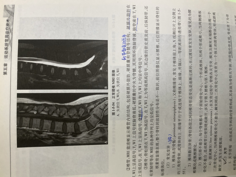
The image shows MRI scans of a normal lumbar spine. Image A is a sagittal T1-weighted image, and image B is a sagittal T2-weighted image. The descriptions highlight the MRI appearance of the spinal canal, including epidural fat (high signal on T1), dura (low signal on T1, high on T2), cerebrospinal fluid (low signal on T1, high on T2), and spinal cord (intermediate signal on both). Vertebral bodies have high signal on T1 (due to bone marrow) and intermediate signal on T2, with low signal cortices and ligaments. Common variations include persistent osteoepiphysis, vertebral number abnormalities, and spina bifida.
LANDLORD242

Dec 24, 2025, 5:09 AM

Chongqing High-tech Zone, China

Stake attention in this memory

medical imaging
MRI
spine
vertebrae
anatomy

The image shows MRI scans of a normal lumbar spine. Image A is a sagittal T1-weighted image, and image B is a sagittal T2-weighted image. The descriptions highlight the MRI appearance of the spinal canal, including epidural fat (high signal on T1), dura (low signal on T1, high on T2), cerebrospinal fluid (low signal on T1, high on T2), and spinal cord (intermediate signal on both). Vertebral bodies have high signal on T1 (due to bone marrow) and intermediate signal on T2, with low signal cortices and ligaments. Common variations include persistent osteoepiphysis, vertebral number abnormalities, and spina bifida.

Loading AttnAds…

transactions
revenues
stakers
Earliest
Latest
Highest stake

No transactions found
More from this user

More from Chongqing High-tech Zone

LANDLORD242

Dec 24, 2025, 5:09 AM

Chongqing High-tech Zone, China

Licensed under Creative Commons Attribution 4.0 International License. You are free to share and adapt this content with proper attribution.