
Stake attention in this memory
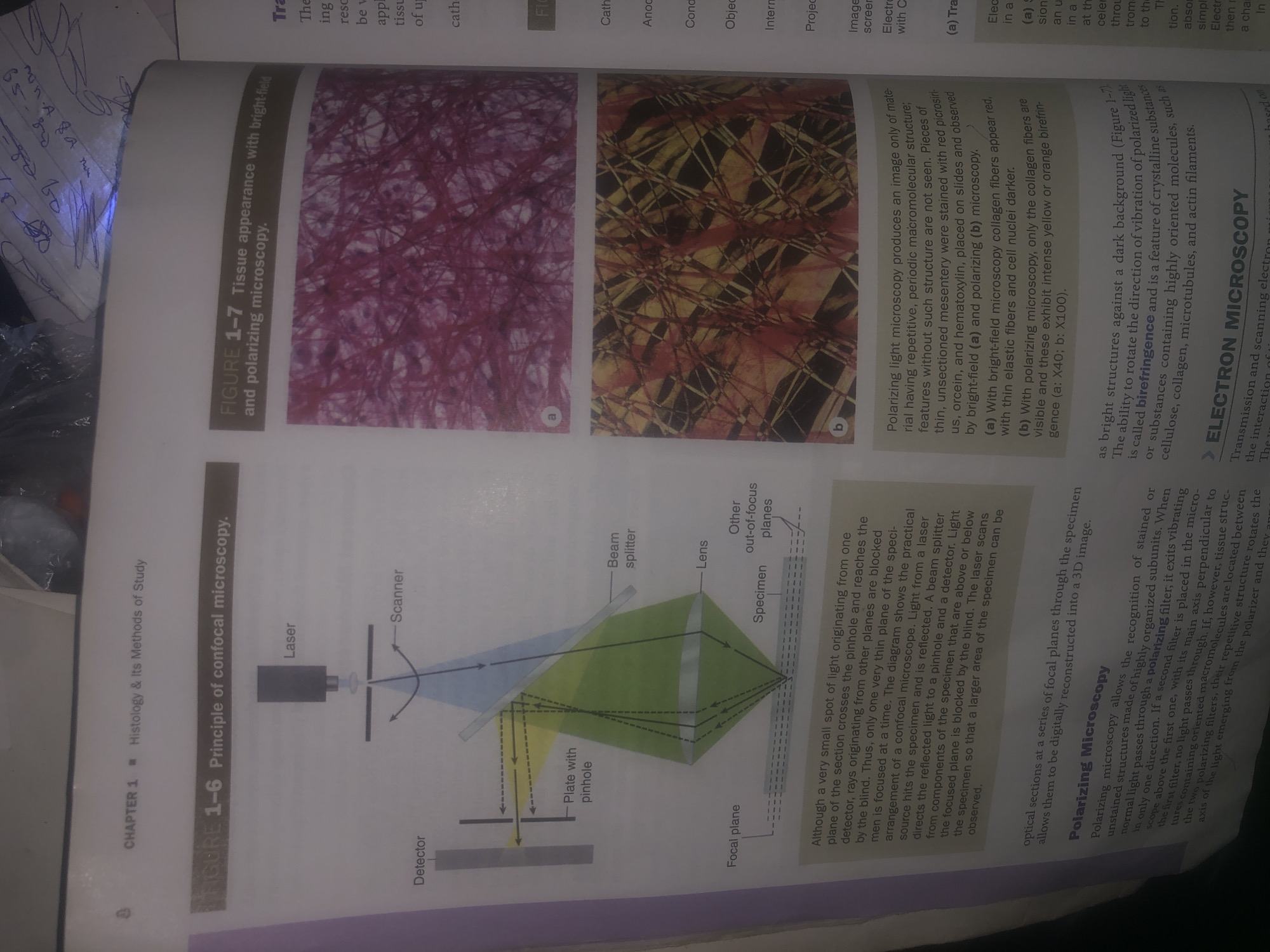
The image displays pages from a textbook discussing microscopy techniques. On the left, Figure 1-6 illustrates the principle of confocal microscopy with a diagram showing a laser, scanner, beam splitter, lens, pinhole, and detector. The accompanying text explains how confocal microscopy works by focusing on a thin focal plane and blocking out-of-focus light. On the right, Figure 1-7 presents tissue appearance under bright-field and polarizing microscopy. Image (a) shows collagen fibers appearing red under bright-field microscopy, with thin elastic fibers and cell nuclei appearing darker. Image (b) shows the same tissue under polarizing microscopy, where the collagen fibers are visible and exhibit intense yellow or orange birefringence. The text describes that polarizing light microscopy produces an image of material having repetitive, periodic macromolecular structure. The ability to rotate the direction of vibration of polarized light, called birefringence, is a feature of crystalline substances or substances containing highly oriented molecules. The text also introduces "Polarizing Microscopy" as a technique for recognizing stained or unstained subunits, and discusses the use of polarizing filters to achieve this. Further down, "ELECTRON MICROSCOPY" is introduced as a technique for transmission and scanning electron microscopy. There is no information within the image or its surrounding context that relates to the city of Wukari, Nigeria. The content is purely academic and scientific.
Loading AttnAds…
No transactions found