Stake attention in this memory
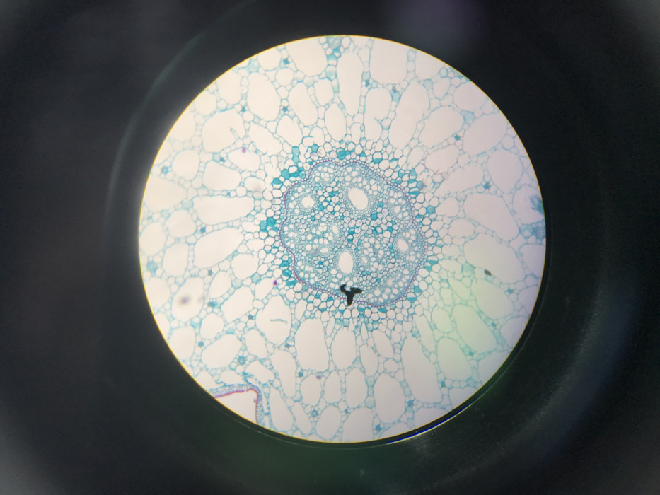
The image displays a microscopic, circular field of view against a dark background, showing a stained cross-section of plant tissue, likely a root or stem. The scene is observed through a microscope at an undisclosed facility in Midrand, South Africa. The primary subject is a plant tissue specimen characterized by a central, roughly circular vascular cylinder composed of densely packed cells stained in varying shades of light and darker blue. Within this core, several larger, clearer circular structures are visible, consistent with xylem vessels. Radiating outwards from the central cylinder are multiple layers of larger, more irregularly shaped cells, predominantly light blue and translucent, which form the cortex. Towards the left periphery of the field, a distinct outermost layer, possibly the epidermis, is present, showing some minute red-stained cellular elements. A prominent, dark, irregularly shaped object, resembling a three-pronged star or a piece of particulate debris, is situated within the cortical region, positioned to the left of the central vascular cylinder. Additionally, several smaller, faint purple or dark specks are scattered throughout the lighter-stained cells. No human figures, tools, or direct actions are visible within the microscopic field.
No transactions found