
Stake attention in this memory
radiology
medical imaging
bone
anatomy
pelvis
x-ray
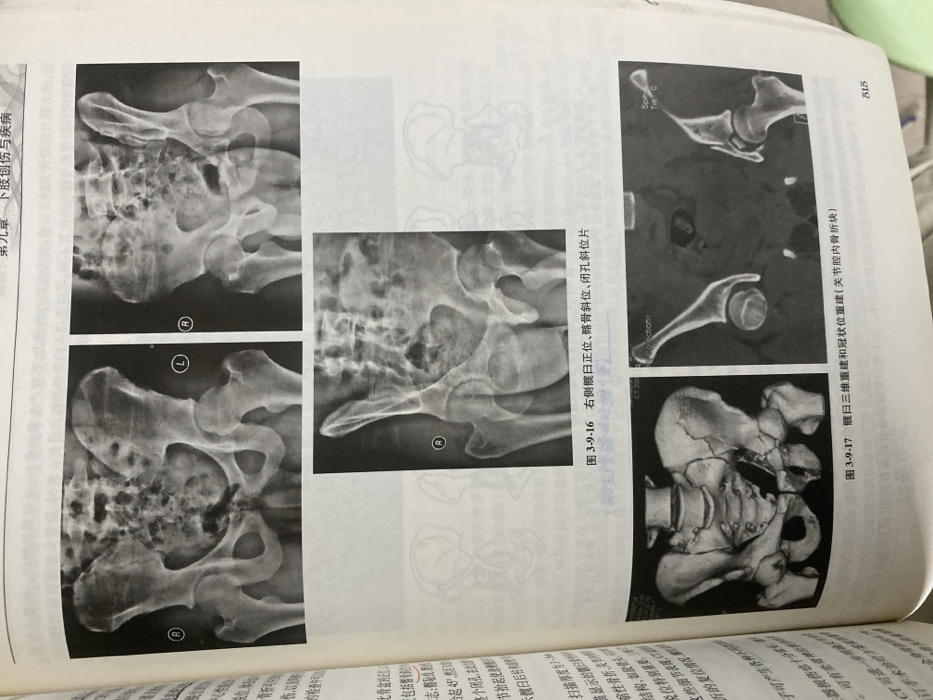
The image shows X-ray and CT scans of a fractured pelvis. The left side displays radiographic images of the right (R) and left (L) hips from different angles, likely demonstrating various views for assessing the fracture. The right side presents a CT scan, including a 3D reconstruction and coronal view, illustrating the location and extent of the fracture within the joint cavity. Text surrounding the images mentions terms like "obturator foramen," "iliac oblique view," "joint cavity fracture," and scanning parameters related to the CT scan.
Loading AttnAds…
transactions
revenues
stakers
Earliest
Latest
Highest stake
No transactions found