
Stake attention in this memory
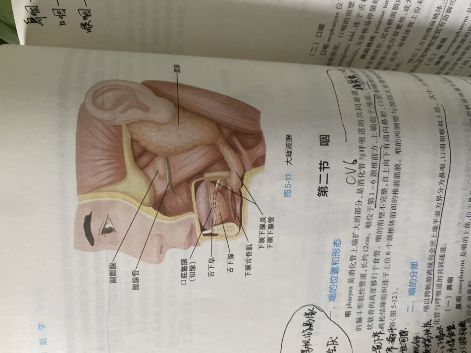
anatomy diagram
human anatomy
medical illustration
Chinese text
textbook
The image depicts an anatomical illustration of the pharynx region. Labels point to structures like the parotid gland, parotid duct, floor of the mouth mucosa, sublingual caruncle, sublingual gland, mylohyoid muscle, submandibular gland, and submandibular duct. The illustration includes the location and morphology of the pharynx, described as a funnel-shaped muscular tube extending from the base of the skull to the esophagus, about 12 cm long. It is situated anterior to the first to sixth cervical vertebrae. The illustration refers to the oropharynx and laryngopharynx.
Loading AttnAds…
transactions
revenues
stakers
Earliest
Latest
Highest stake
No transactions found