Sign in
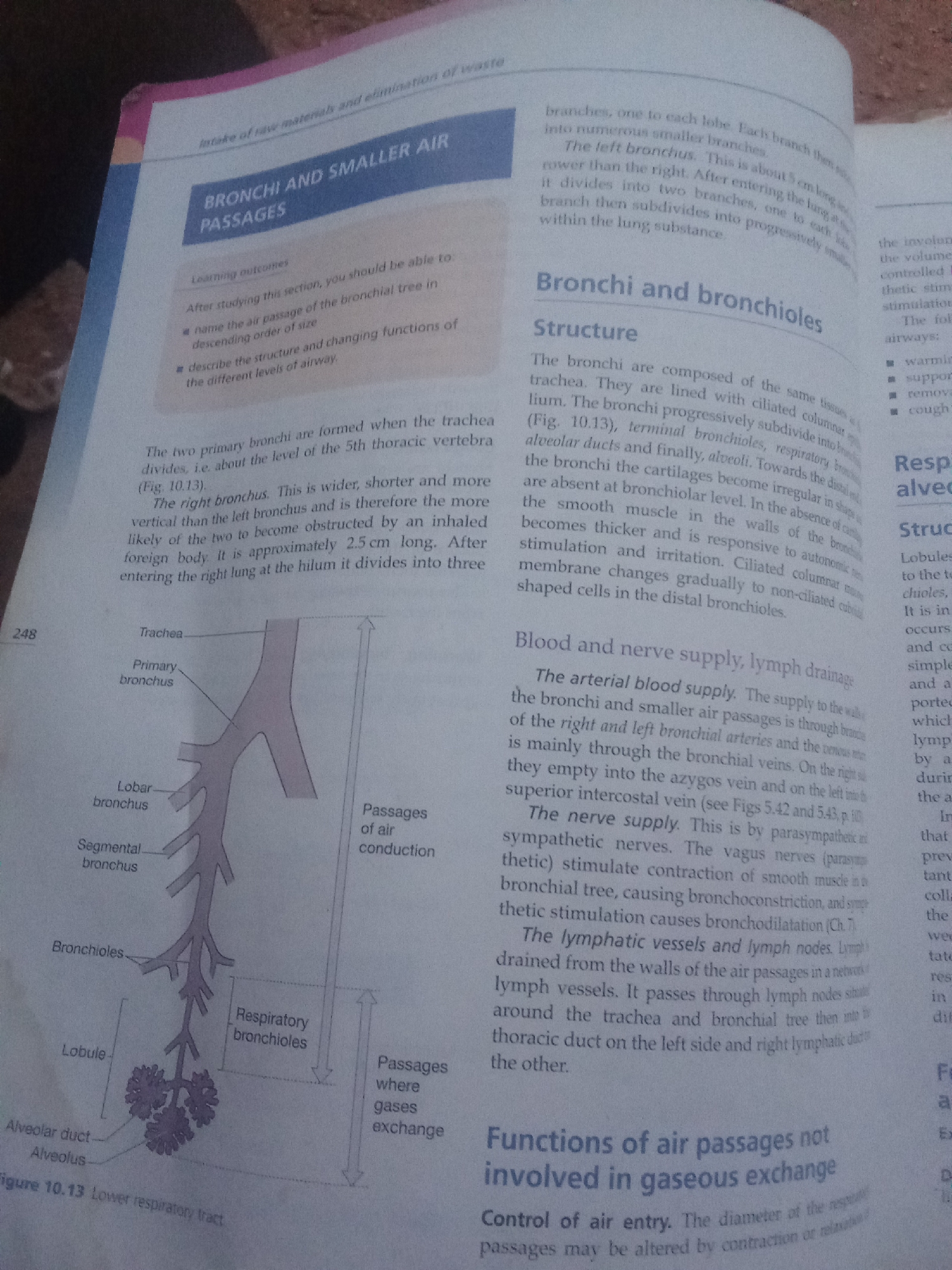
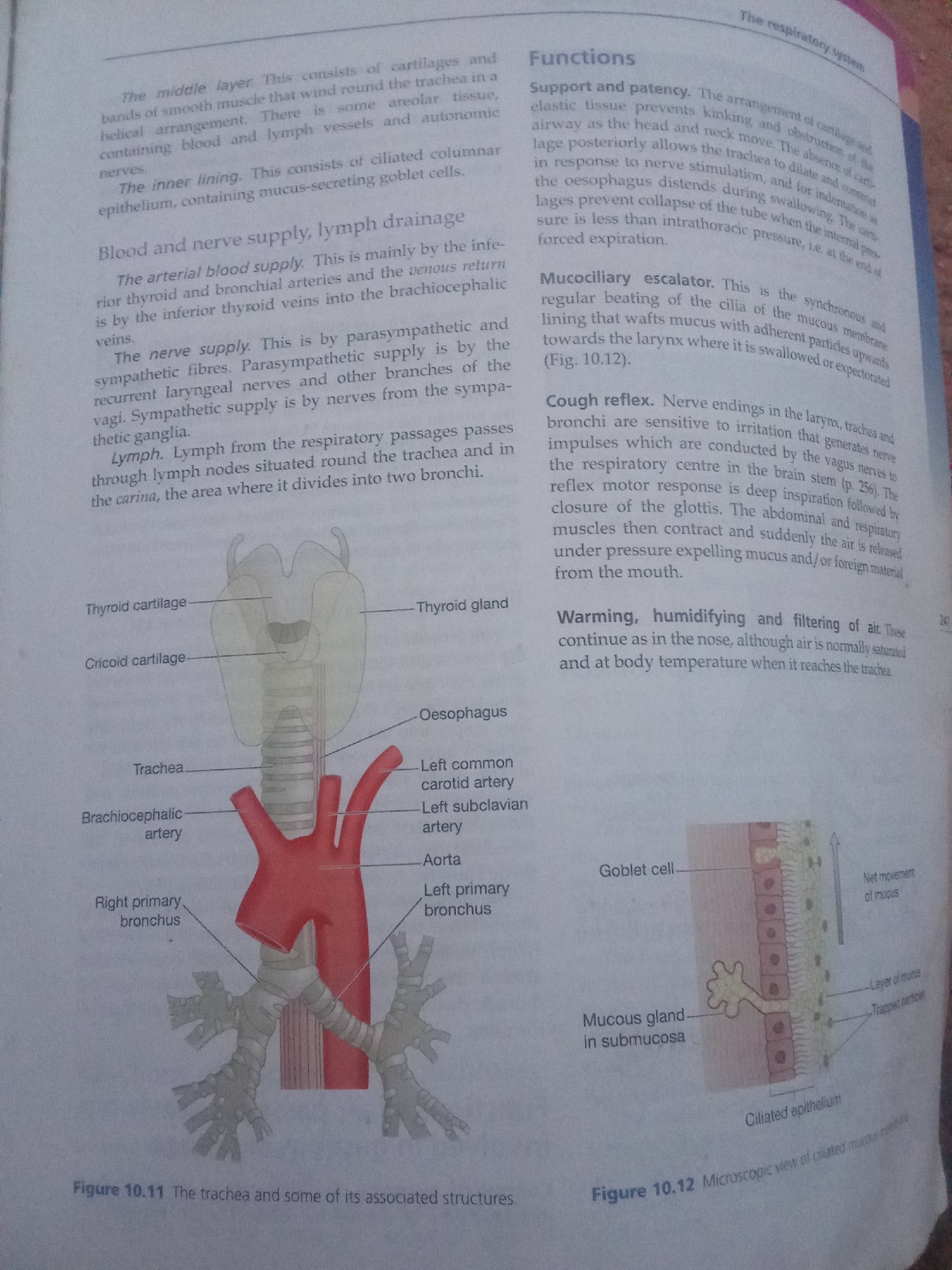
This image is a scanned page from an anatomy textbook, not a photograph of a real-world location. Therefore, I cannot provide any details about Jalingo, Nigeria.

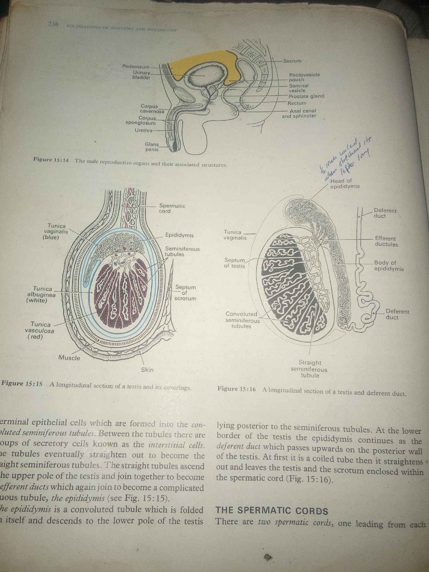
The image contains two diagrams illustrating the male reproductive system.

Figure 15:14 shows a sagittal view of the male pelvic region, depicting organs such as the peritoneum, urinary bladder, rectum, seminal vesicle, prostate gland, and penis.

Figure 15:15 and 15:16 provide a detailed longitudinal section of a testis and its coverings, as well as the epididymis and deferent duct. Various anatomical structures within the testis, including seminiferous tubules, septa, and tunics, are labeled. The surrounding text describes the structure and function of the seminiferous tubules, interstitial cells, epididymis, and deferent duct, as well as the composition of the spermatic cord.
FM-KpyEx1

Jun 30, 2026, 4:02 PM

Jalingo, Nigeria

Stake attention in this memory

informative
clinical
academic
analytical
neutral

This image is a scanned page from an anatomy textbook, not a photograph of a real-world location. Therefore, I cannot provide any details about Jalingo, Nigeria. The image contains two diagrams illustrating the male reproductive system. Figure 15:14 shows a sagittal view of the male pelvic region, depicting organs such as the peritoneum, urinary bladder, rectum, seminal vesicle, prostate gland, and penis. Figure 15:15 and 15:16 provide a detailed longitudinal section of a testis and its coverings, as well as the epididymis and deferent duct. Various anatomical structures within the testis, including seminiferous tubules, septa, and tunics, are labeled. The surrounding text describes the structure and function of the seminiferous tubules, interstitial cells, epididymis, and deferent duct, as well as the composition of the spermatic cord.

Loading AttnAds…

transactions
revenues
stakers
Earliest
Latest
Highest stake

No transactions found

More from this user

More from Jalingo

FM-KpyEx1

Jun 30, 2026, 4:02 PM

Jalingo, Nigeria

Licensed under Creative Commons Attribution 4.0 International License. You are free to share and adapt this content with proper attribution.