
Sign in
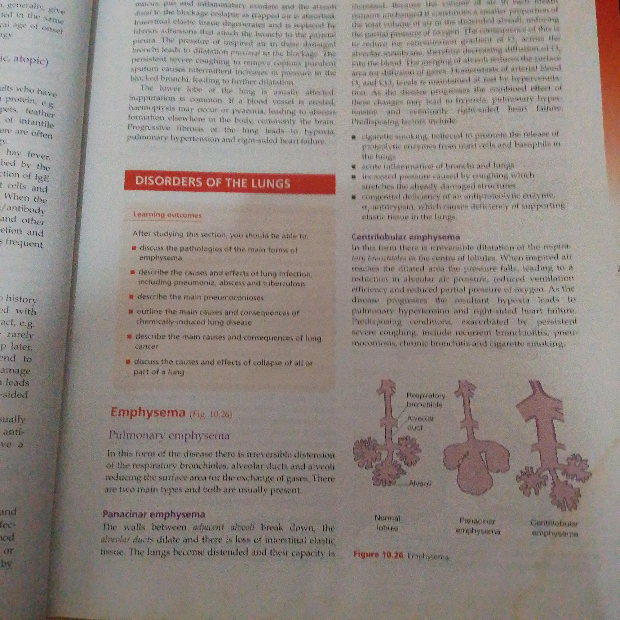
This is a close-up, overhead photograph of an open textbook, focusing on a page detailing medical information about "Disorders of the Lungs," specifically "Emphysema." The text is printed in black ink on white paper.

The page is divided into several sections. At the top, there is a heading "DISORDERS OF THE LUNGS" in a red bar, followed by "Learning outcomes" which lists several bullet points detailing what a student should be able to do after studying the section. Below this, the primary topic "Emphysema" is introduced with a figure reference "(Fig. 10.26)". The text then elaborates on "Pulmonary emphysema" and its two main types: "Panacinar emphysema" and "Centrilobular emphysema."

To the right of the text, a diagram labeled "Figure 10.26 Emphysema" illustrates the structures of a normal lobule, panacinar emphysema, and centrilobular emphysema. These diagrams show simplified representations of bronchioles, alveolar ducts, and alveoli, highlighting the changes that occur in emphysema.

On the left side of the page, there are also several paragraphs of text that appear to be continuations or related information from previous sections, discussing conditions like bronchitis and atopic reactions, and mentioning symptoms like hay fever and IgE.

The lighting suggests indoor illumination, typical of reading in a well-lit room. There are no people visible in the image. The focus is entirely on the printed content of the textbook. The overall impression is one of educational material being presented.
FM-66btC3

Jun 30, 2026, 1:33 PM

Jalingo, Nigeria

Stake attention in this memory

informative
educational
medical
scientific
clinical

This is a close-up, overhead photograph of an open textbook, focusing on a page detailing medical information about "Disorders of the Lungs," specifically "Emphysema." The text is printed in black ink on white paper. The page is divided into several sections. At the top, there is a heading "DISORDERS OF THE LUNGS" in a red bar, followed by "Learning outcomes" which lists several bullet points detailing what a student should be able to do after studying the section. Below this, the primary topic "Emphysema" is introduced with a figure reference "(Fig. 10.26)". The text then elaborates on "Pulmonary emphysema" and its two main types: "Panacinar emphysema" and "Centrilobular emphysema." To the right of the text, a diagram labeled "Figure 10.26 Emphysema" illustrates the structures of a normal lobule, panacinar emphysema, and centrilobular emphysema. These diagrams show simplified representations of bronchioles, alveolar ducts, and alveoli, highlighting the changes that occur in emphysema. On the left side of the page, there are also several paragraphs of text that appear to be continuations or related information from previous sections, discussing conditions like bronchitis and atopic reactions, and mentioning symptoms like hay fever and IgE. The lighting suggests indoor illumination, typical of reading in a well-lit room. There are no people visible in the image. The focus is entirely on the printed content of the textbook. The overall impression is one of educational material being presented.

Loading AttnAds…

transactions
revenues
stakers
Earliest
Latest
Highest stake

No transactions found

More from this user

More from Jalingo

FM-66btC3

Jun 30, 2026, 1:33 PM

Jalingo, Nigeria

Licensed under Creative Commons Attribution 4.0 International License. You are free to share and adapt this content with proper attribution.