Oct 27, 2025
Stake attention in this memory
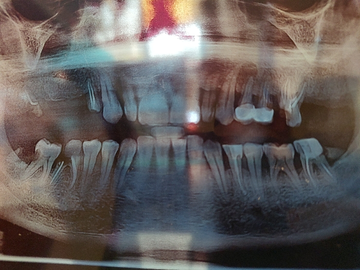
A panoramic dental radiograph (orthopantomogram) displays the human dentition, maxillary, and mandibular bone structures. The image reveals numerous missing teeth, particularly in the posterior regions of both the upper and lower arches. Existing dentition shows evidence of multiple radiopaque dental restorations consistent with fillings and possibly crowns or prosthetic work. Visible tooth roots are present for the remaining teeth. The surrounding bone anatomy of the maxilla and mandible is discernible, including portions of the maxillary sinuses and temporomandibular joint areas. The image itself exhibits glare and reflections, suggesting it is a photograph of a physical X-ray film or digital screen. The location is Unknown, Unknown.
No transactions found
More from this user