
Sign in
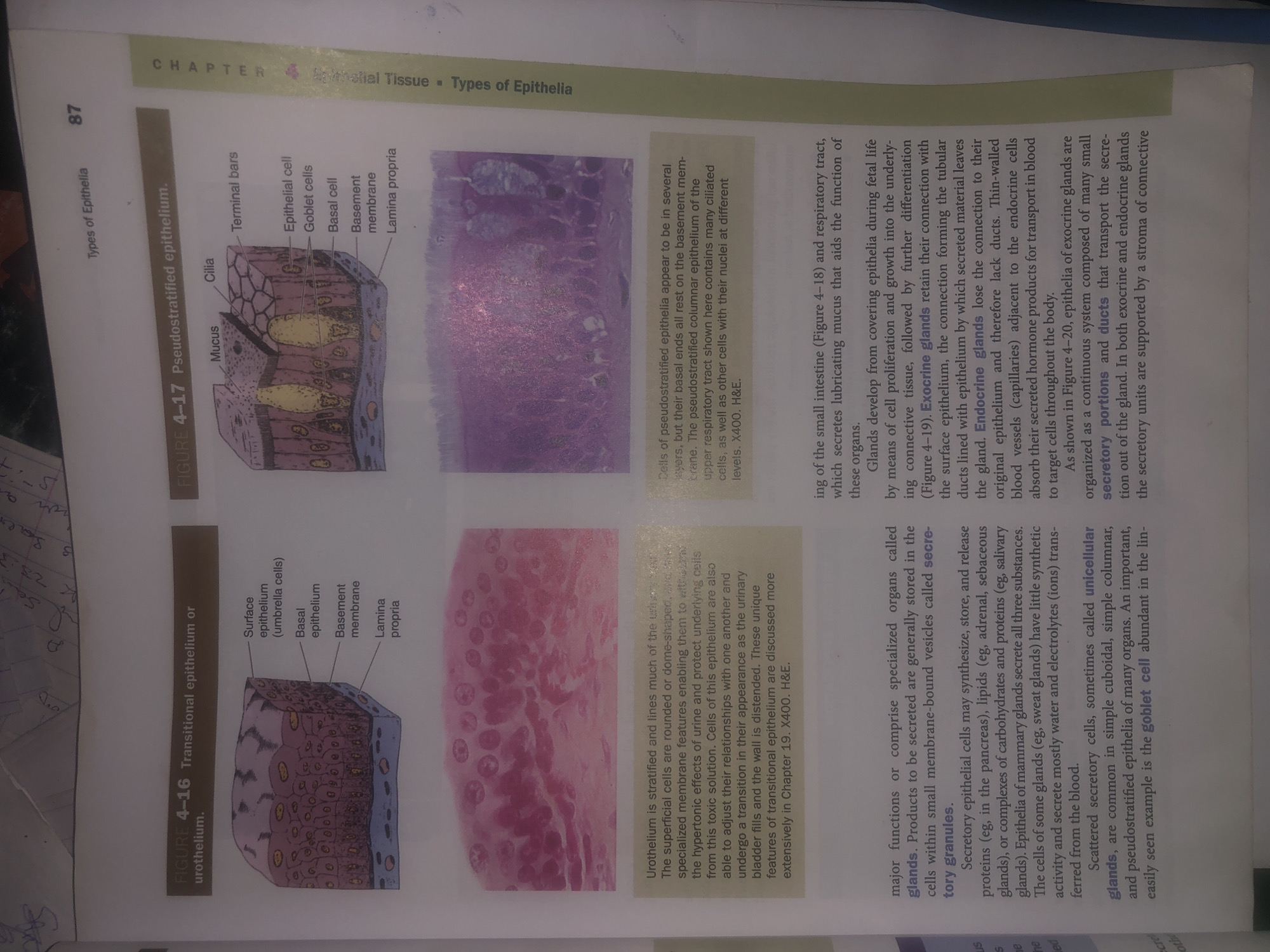
The image displays pages from a textbook illustrating different types of epithelial tissues.

On the left side, Figure 4-16 shows a diagram and microscopic view of transitional epithelium or urothelium, highlighting its stratified nature with surface epithelium (umbrella cells), basal epithelium, basement membrane, and lamina propria. The accompanying text describes urothelium lining the urinary tract, its ability to distend, and its protective function against urine.

On the right side, Figure 4-17 presents a diagram and microscopic view of pseudostratified epithelium, showing mucus, cilia, terminal bars, epithelial cells, goblet cells, basal cell, basement membrane, and lamina propria. The text explains that pseudostratified epithelia appear stratified but are not, with basal ends resting on the basement membrane. It specifically mentions pseudostratified columnar epithelium of the upper respiratory tract, which contains ciliated cells and others with nuclei at different levels.

Below these figures, there is a discussion about glands, their development from epithelia, and the distinction between exocrine glands (retaining connection to the surface epithelium via ducts) and endocrine glands (losing this connection and secreting hormones directly into blood vessels). It also touches upon the organization of exocrine glands into secretory portions and ducts.

The image does not contain any information related to the city of Wukari, Nigeria.
Bigstev

Jun 12, 2026, 1:55 AM

Wukari, Nigeria

Short Term

Stake attention in this memory

informative
educational
scientific
detailed
anatomical

The image displays pages from a textbook illustrating different types of epithelial tissues. On the left side, Figure 4-16 shows a diagram and microscopic view of transitional epithelium or urothelium, highlighting its stratified nature with surface epithelium (umbrella cells), basal epithelium, basement membrane, and lamina propria. The accompanying text describes urothelium lining the urinary tract, its ability to distend, and its protective function against urine. On the right side, Figure 4-17 presents a diagram and microscopic view of pseudostratified epithelium, showing mucus, cilia, terminal bars, epithelial cells, goblet cells, basal cell, basement membrane, and lamina propria. The text explains that pseudostratified epithelia appear stratified but are not, with basal ends resting on the basement membrane. It specifically mentions pseudostratified columnar epithelium of the upper respiratory tract, which contains ciliated cells and others with nuclei at different levels. Below these figures, there is a discussion about glands, their development from epithelia, and the distinction between exocrine glands (retaining connection to the surface epithelium via ducts) and endocrine glands (losing this connection and secreting hormones directly into blood vessels). It also touches upon the organization of exocrine glands into secretory portions and ducts. The image does not contain any information related to the city of Wukari, Nigeria.

Symbol

B6EA5

Volume

11,550

Creator

+$0.03

Revenue

+$0.05

TVL

$2.95

1D
1W
1M
All

Loading AttnAds…

transactions
revenues
stakers
Earliest
Latest
Highest stake

1

ZaddyTanko

Staked 26d ago

$2.93 USD

+$0.02 USD

+0.7%

2

Rev Bot 🤖💰

Injected revenue 26d ago

Revenue bonus for the last stake.

+$0.06 USD

More from this user

More from Wukari

Bigstev

Jun 12, 2026, 1:55 AM

Wukari, Nigeria

Licensed under Creative Commons Attribution 4.0 International License. You are free to share and adapt this content with proper attribution.