
Stake attention in this memory
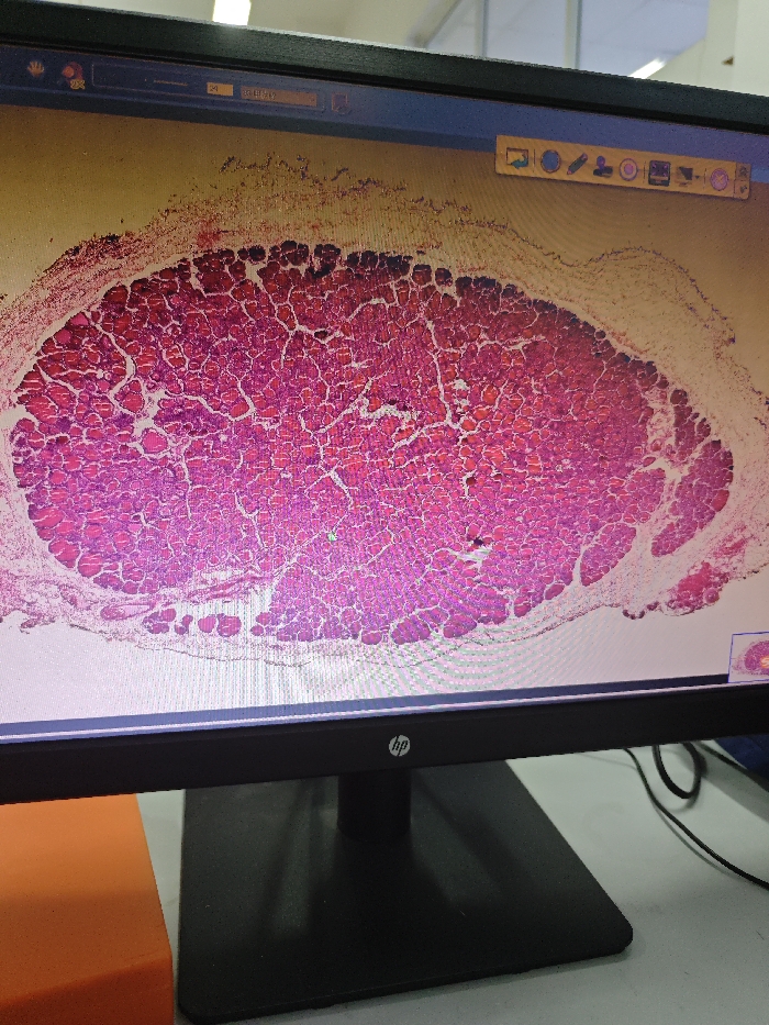
An HP monitor displays a magnified histological image of biological tissue, characterized by densely packed cells with prominent pink/red cytoplasm and darker nuclei, typical of H&E stained muscle or glandular tissue. The tissue section is irregularly oval-shaped against a light yellow digital background. User interface elements are visible at the top of the screen, including application icons, a progress bar, Chinese text reading "20 样本1912" (20 Sample 1912), and a toolbar containing various image manipulation and annotation tools. A small green dot, likely a cursor, is present near the center of the tissue image, and a thumbnail of the entire tissue section is in the lower right corner of the digital display. The monitor features a black bezel and a rectangular black stand. It rests on a light-colored surface, possibly a laboratory bench or desk. An orange object, possibly a foam block, is positioned to the left of the monitor stand, and a black cable is visible on the right. The background behind the monitor is blurred, suggesting an indoor environment, likely a laboratory or research facility, located in the Chongqing High-tech Zone, China. No individuals are visible.
Loading AttnAds…
No transactions found