
Sign in
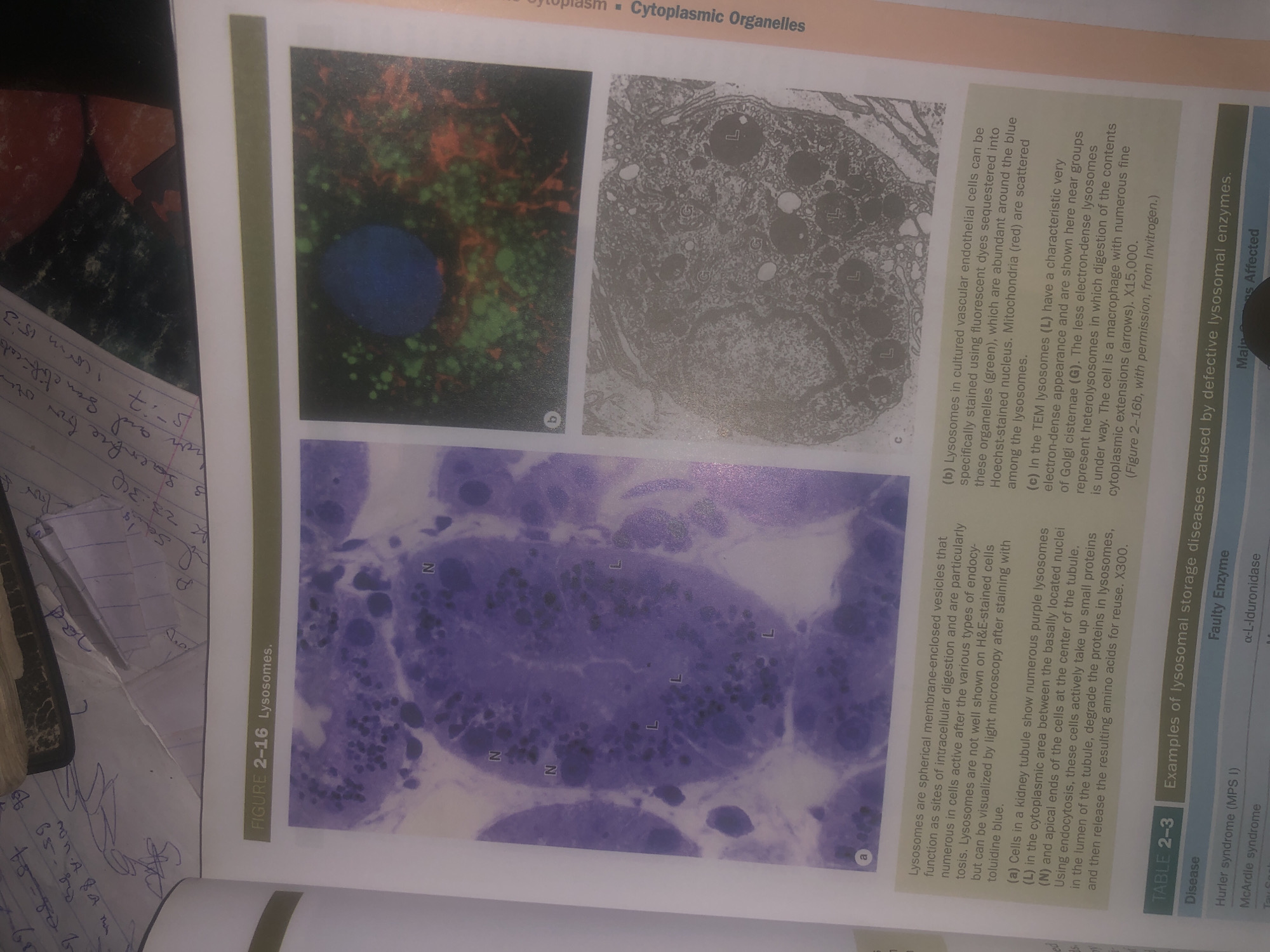
The image displays a page from a textbook or scientific publication detailing lysosomes. It features Figure 2-16 with three micrographs labeled a, b, and c. Micrograph (a) shows cells with purple structures labeled 'N' and 'L', described as lysosomes in a kidney tubule. Micrograph (b) is a fluorescence micrograph of lysosomes in cultured vascular endothelial cells, with green and red staining indicating specific cellular components. Micrograph (c) is a transmission electron micrograph of lysosomes (labeled 'L') and Golgi cisternae (labeled 'G') within a cell, possibly a macrophage, showing cytoplasmic extensions. The accompanying text provides descriptions of these micrographs and defines lysosomes as membrane-enclosed vesicles involved in intracellular digestion. Below the micrographs is Table 2-3, which lists examples of lysosomal storage diseases caused by defective lysosomal enzymes, with columns for Disease, Faulty Enzyme, and Main Tissues Affected. The image content is purely scientific and educational, with no discernible connection to the city of Wukari, Nigeria, or any forensic context.
Bigstev

Jun 12, 2026, 1:42 AM

Wukari, Nigeria

Moment

Stake attention in this memory

curiosity
discovery
scientific
educational
analytical

The image displays a page from a textbook or scientific publication detailing lysosomes. It features Figure 2-16 with three micrographs labeled a, b, and c. Micrograph (a) shows cells with purple structures labeled 'N' and 'L', described as lysosomes in a kidney tubule. Micrograph (b) is a fluorescence micrograph of lysosomes in cultured vascular endothelial cells, with green and red staining indicating specific cellular components. Micrograph (c) is a transmission electron micrograph of lysosomes (labeled 'L') and Golgi cisternae (labeled 'G') within a cell, possibly a macrophage, showing cytoplasmic extensions. The accompanying text provides descriptions of these micrographs and defines lysosomes as membrane-enclosed vesicles involved in intracellular digestion. Below the micrographs is Table 2-3, which lists examples of lysosomal storage diseases caused by defective lysosomal enzymes, with columns for Disease, Faulty Enzyme, and Main Tissues Affected. The image content is purely scientific and educational, with no discernible connection to the city of Wukari, Nigeria, or any forensic context.

Symbol

BDBEE

Volume

11,269

Creator

+$0.03

Revenue

+$0.06

TVL

$2.81

1D
1W
1M
All

Loading AttnAds…

transactions
revenues
stakers
Earliest
Latest
Highest stake

1

ZaddyTanko

Staked 26d ago

$2.78 USD

+$0.03 USD

+1%

2

Rev Bot 🤖💰

Injected revenue 26d ago

Revenue bonus for the last stake.

+$0.07 USD

More from this user

More from Wukari

Bigstev

Jun 12, 2026, 1:42 AM

Wukari, Nigeria

Licensed under Creative Commons Attribution 4.0 International License. You are free to share and adapt this content with proper attribution.