Stake attention in this memory
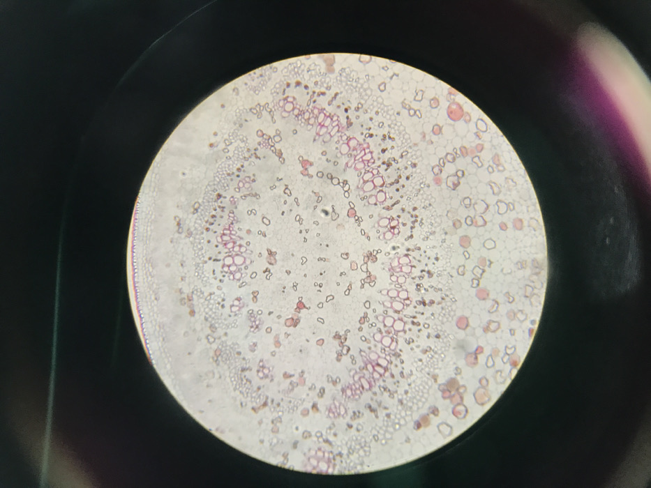
The image displays a circular, magnified view of a stained biological specimen, consistent with a transverse section of plant tissue, observed through a microscope. The specimen exhibits distinct cellular structures including an outer epidermal layer, a cortex, and multiple vascular bundles arranged in a ring towards the center. These vascular bundles appear stained purple and pink, highlighting their cellular components. The ground tissue, composed of various parenchyma cells, fills the internal regions. Scattered red and pink spherical and irregular structures are visible throughout the section. The immediate background outside the circular field of view is dark. This microscopic observation occurred in Midrand, South Africa. No individuals, actions, or direct interactions are depicted. The environment depicted is solely the microscopic field.
No transactions found