
Sign in
The image displays anatomical diagrams of the human eye and its accessory structures, specifically focusing on the lacrimal apparatus. There is no information about Jalingo, Nigeria.

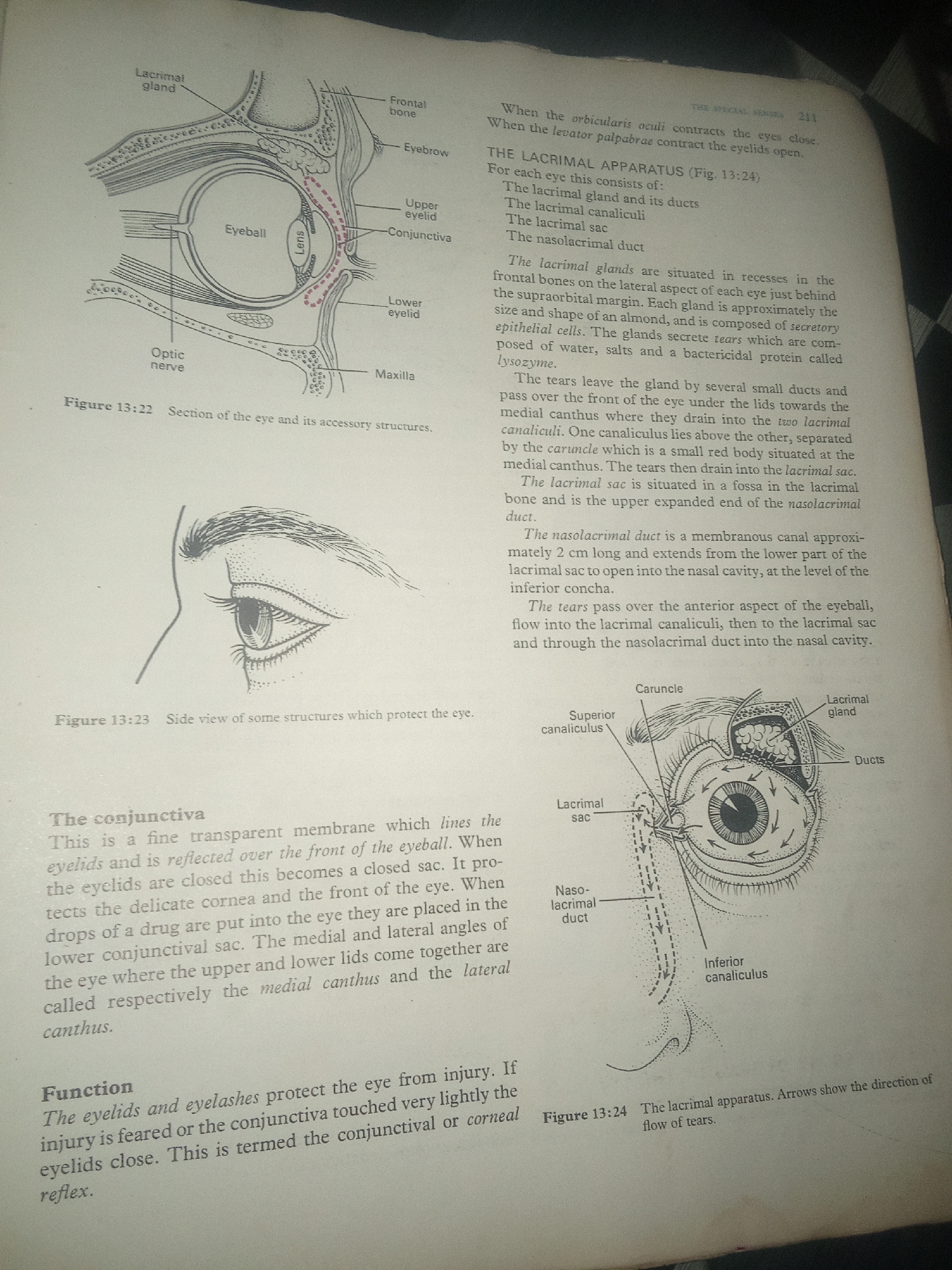
The first diagram (Figure 13:22) is a sagittal section of the eye, illustrating the eyeball, lens, optic nerve, lacrimal gland, frontal bone, eyebrow, upper eyelid, lower eyelid, and maxilla. The conjunctiva is also depicted lining the eyelids and reflecting over the front of the eyeball.

The second diagram (Figure 13:23) shows a side view of the structures protecting the eye, including the eyebrow and eyelids.

The third and largest diagram (Figure 13:24) details the lacrimal apparatus. It shows the lacrimal gland, its ducts, the superior and inferior canaliculi, the lacrimal sac, and the nasolacrimal duct. Arrows indicate the flow of tears from the lacrimal gland across the surface of the eye, into the canaliculi, then to the lacrimal sac, and finally down the nasolacrimal duct into the nasal cavity.

Accompanying text describes the function of the lacrimal apparatus in producing and draining tears, and also discusses the conjunctiva.
FM-KpyEx1

Jun 30, 2026, 3:57 PM

Jalingo, Nigeria

Stake attention in this memory

informative
educational
medical
anatomical
detailed

The image displays anatomical diagrams of the human eye and its accessory structures, specifically focusing on the lacrimal apparatus. There is no information about Jalingo, Nigeria. The first diagram (Figure 13:22) is a sagittal section of the eye, illustrating the eyeball, lens, optic nerve, lacrimal gland, frontal bone, eyebrow, upper eyelid, lower eyelid, and maxilla. The conjunctiva is also depicted lining the eyelids and reflecting over the front of the eyeball. The second diagram (Figure 13:23) shows a side view of the structures protecting the eye, including the eyebrow and eyelids. The third and largest diagram (Figure 13:24) details the lacrimal apparatus. It shows the lacrimal gland, its ducts, the superior and inferior canaliculi, the lacrimal sac, and the nasolacrimal duct. Arrows indicate the flow of tears from the lacrimal gland across the surface of the eye, into the canaliculi, then to the lacrimal sac, and finally down the nasolacrimal duct into the nasal cavity. Accompanying text describes the function of the lacrimal apparatus in producing and draining tears, and also discusses the conjunctiva.

Loading AttnAds…

transactions
revenues
stakers
Earliest
Latest
Highest stake

No transactions found

More from this user

More from Jalingo

FM-KpyEx1

Jun 30, 2026, 3:57 PM

Jalingo, Nigeria

Licensed under Creative Commons Attribution 4.0 International License. You are free to share and adapt this content with proper attribution.