Stake attention in this memory
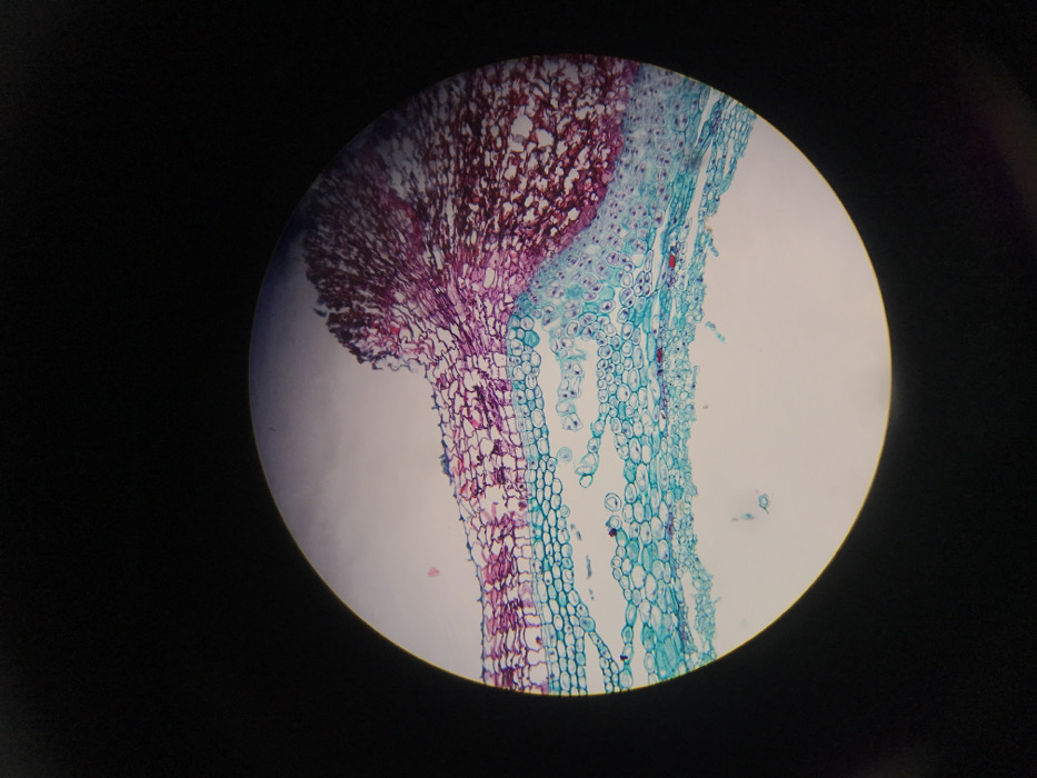
The image displays a microscopic cross-section of biological tissue, observed within a circular field of view, against a black background. The location is Midrand, South Africa, as provided. The tissue exhibits distinct differential staining, revealing two primary cellular regions. The upper and right portions of the field are dominated by cells stained in shades of magenta and deep red. These cells appear densely packed and form an irregular, somewhat convoluted mass, particularly concentrated in the upper-right quadrant. Below this, a distinct layer of cells is stained in cyan and blue-green hues. These cyan-stained cells are arranged in more organized layers, with individual cells appearing round or oval, and some featuring clearer internal structures or lumens. A clear, irregular boundary separates the magenta and cyan cellular populations. No individuals or active interactions are visible. The subject is a static biological specimen under magnification. The objects are stained cells and tissue structures. The environment depicted is solely the microscopic field, implying examination in a laboratory setting.
No transactions found