
Stake attention in this memory
histology
esophagus
stomach
anatomical
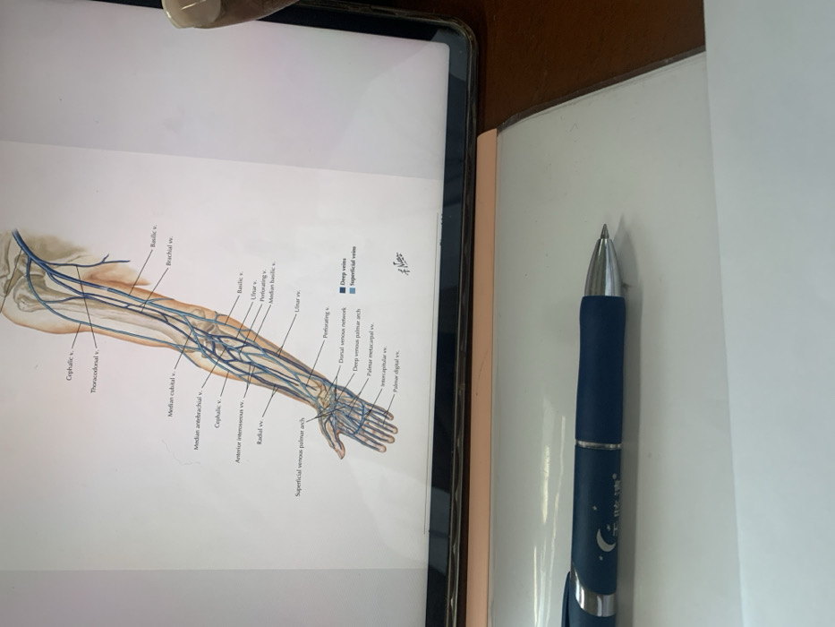
medical illustration
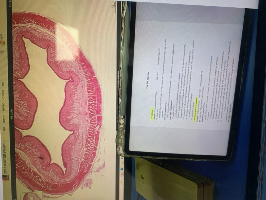
The image shows histological slides and notes from a "5th practice" lab session. One slide appears to be a cross-section of the esophagus, stained with H&E. The notes on the tablet describe the esophagus's layers, including epithelium (purple-blue layer), tunica submucosa (light stained), and muscularis (pink). Under higher magnification, the tunica propria, submucosa (containing lymphoid tissue, blood vessels, and esophageal glands), and muscularis are visible. The muscularis includes longitudinal and circular smooth muscle. Another slide shows a section of the stomach.
Loading AttnAds…
transactions
revenues
stakers
Earliest
Latest
Highest stake
No transactions found