Stake attention in this memory
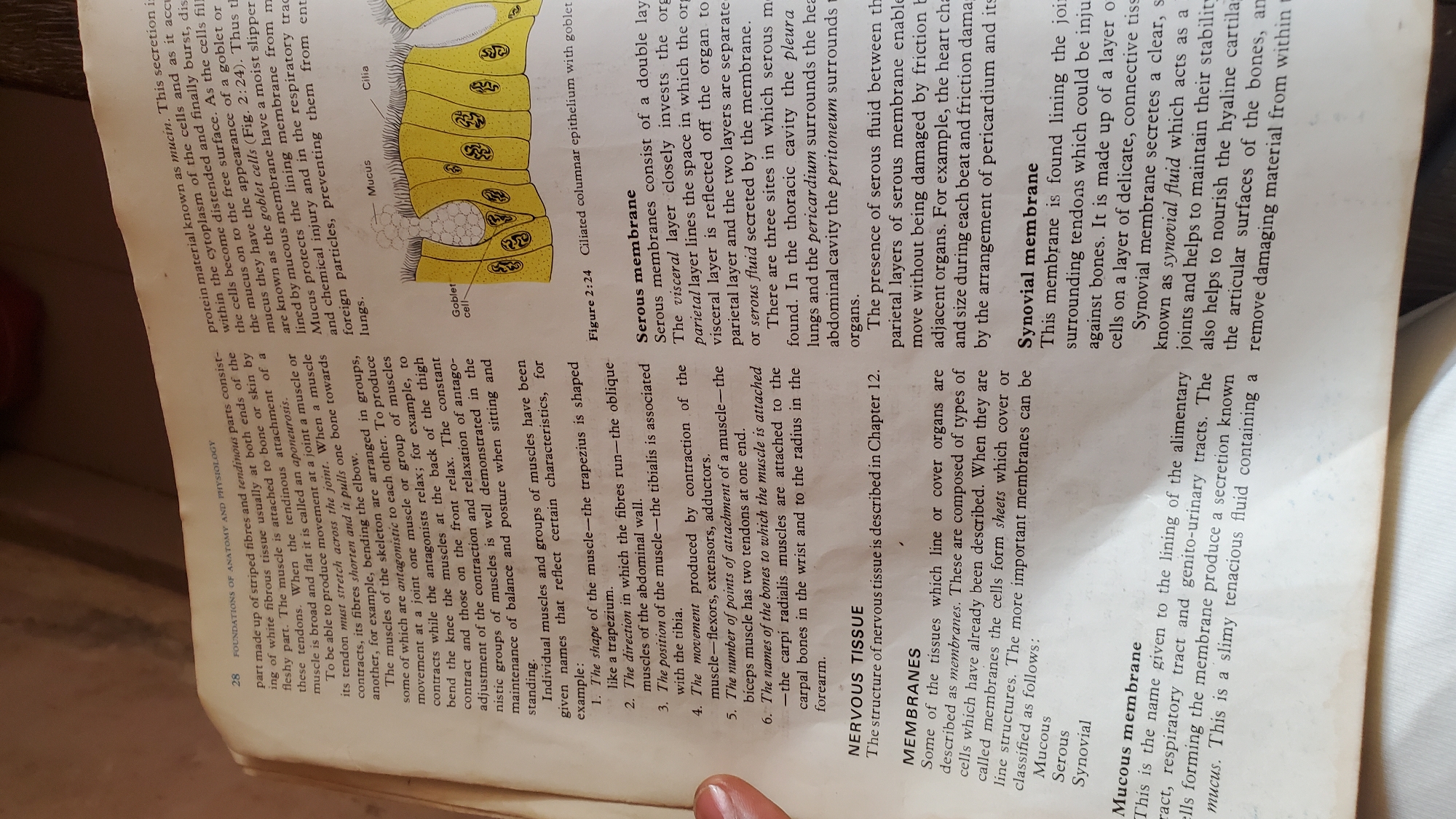
The image displays a page from a textbook, likely related to anatomy and physiology. The page is dominated by an illustration of a biological structure, possibly an epithelial lining with cilia and goblet cells, accompanied by detailed text. The text is divided into sections discussing different types of membranes: mucous, serous, and synovial. There are also sections on muscle and nervous tissue. The illustration features a magnified view of cells, showing cilia projecting from the surface and goblet cells embedded within the epithelium. The cells are depicted in a vibrant yellow, with a darker yellow border and a lighter yellow background. The text is printed in black on a white background. Several labels are present, including "Mucus", "Goblet cell", and "Cilia", pointing to specific parts of the illustration. The illustration is identified as "Figure 2:24 Ciliated columnar epithelium with goblet". The surrounding text provides detailed explanations of the functions and locations of these biological tissues. It describes the composition, secretion, and protective roles of mucous membranes, the structure and locations of serous membranes (pleura, pericardium, peritoneum), and the function of synovial membranes in joints. The text also includes a numbered list of ways muscles are named, referencing examples like the trapezius and biceps. A page number "28" and a chapter heading "FOUNDATIONS OF ANATOMY AND PHYSIOLOGY" are visible at the top of the page. The overall impression is an educational resource aimed at explaining biological concepts through diagrams and textual descriptions.
No transactions found