Sign in
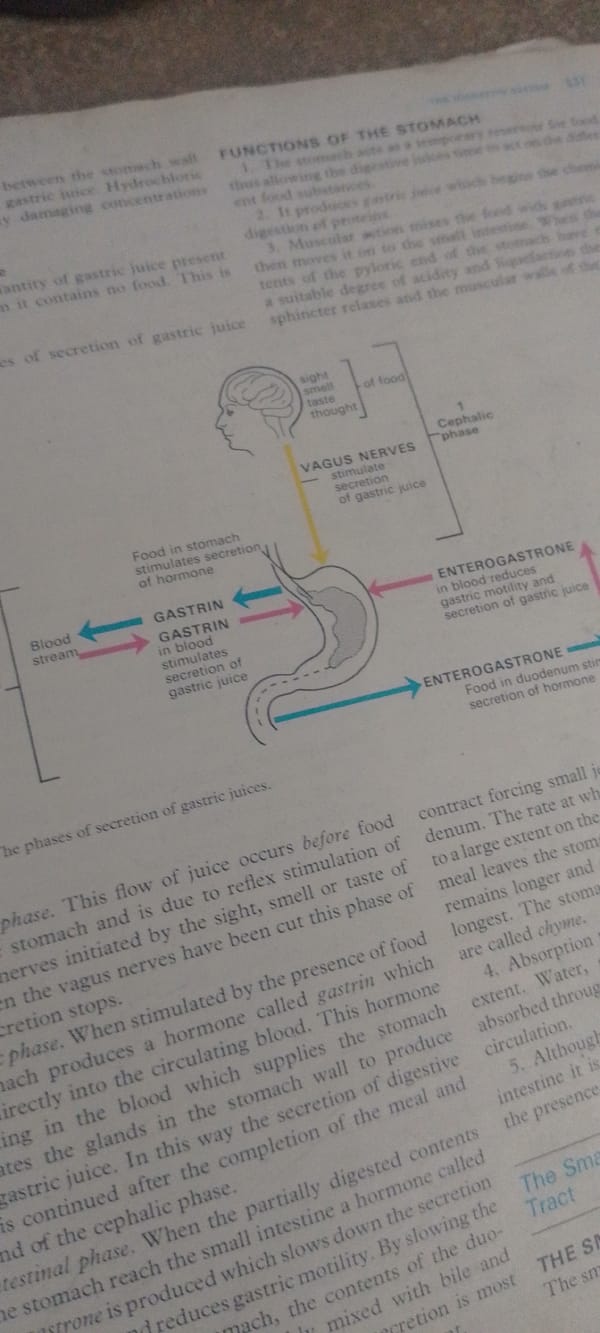
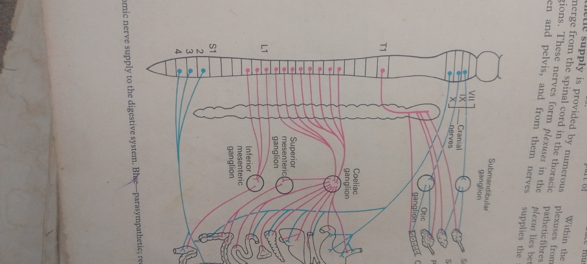
This is a diagram illustrating the autonomic nerve supply to the digestive system, with a focus on the thoracic and pelvic regions. The diagram depicts the spinal cord, with segments labeled S1, L1, and T1. From these segments, various nerves branch out, shown in pink for sympathetic and blue for parasympathetic pathways. These nerves form plexuses, with key ganglia like the superior mesenteric, inferior mesenteric, coeliac, submandibular, and otic ganglia clearly labeled. The diagram also shows a representation of the digestive organs, including the intestines and stomach, indicating how they are innervated.

Visible text includes labels for spinal cord segments (S1, L1, T1, 2, 3, 4), nerve pathways (Cranial nerves), ganglia (Superior mesenteric ganglion, Inferior mesenteric ganglion, Coeliac ganglion, Submandibular ganglion, Otic ganglion), and descriptive text explaining the origin and function of the nerve supply, stating "Autonomic nerve supply to the digestive system. Blue-parasympathetic; red-sympathetic". The text also mentions "plexuses from thoracic", "plexuses in the...", "plexus lies bet", and "supplies the".

The diagram itself is the primary subject, representing a biological and anatomical concept. There are no people, animals, or specific events depicted. The setting is implied to be within an educational or scientific context, such as a textbook or lecture material, due to the nature of the diagram. There are no cues regarding time of day, weather, or emotions.
FM-2L6Lq2

Jun 30, 2026, 5:10 PM

Jauro Yinu, Nigeria

Moment

Stake attention in this memory

educational
scientific
informative
anatomical
diagrammatic

This is a diagram illustrating the autonomic nerve supply to the digestive system, with a focus on the thoracic and pelvic regions. The diagram depicts the spinal cord, with segments labeled S1, L1, and T1. From these segments, various nerves branch out, shown in pink for sympathetic and blue for parasympathetic pathways. These nerves form plexuses, with key ganglia like the superior mesenteric, inferior mesenteric, coeliac, submandibular, and otic ganglia clearly labeled. The diagram also shows a representation of the digestive organs, including the intestines and stomach, indicating how they are innervated. Visible text includes labels for spinal cord segments (S1, L1, T1, 2, 3, 4), nerve pathways (Cranial nerves), ganglia (Superior mesenteric ganglion, Inferior mesenteric ganglion, Coeliac ganglion, Submandibular ganglion, Otic ganglion), and descriptive text explaining the origin and function of the nerve supply, stating "Autonomic nerve supply to the digestive system. Blue-parasympathetic; red-sympathetic". The text also mentions "plexuses from thoracic", "plexuses in the...", "plexus lies bet", and "supplies the". The diagram itself is the primary subject, representing a biological and anatomical concept. There are no people, animals, or specific events depicted. The setting is implied to be within an educational or scientific context, such as a textbook or lecture material, due to the nature of the diagram. There are no cues regarding time of day, weather, or emotions.

Symbol

CFF4A

Volume

2,726

Creator

+$0.00

Revenue

+$0.00

TVL

$0.21

1D
1W
1M
All

Loading AttnAds…

transactions
revenues
stakers
Earliest
Latest
Highest stake

1

FM-FSRgn1

Staked 16d ago

$0.21 USD

+$0.00 USD

+0.8%

2

Rev Bot 🤖💰

Injected revenue 16d ago

Revenue bonus for the last stake.

+$0.00 USD

More from this user

More from Jauro Yinu

FM-2L6Lq2

Jun 30, 2026, 5:10 PM

Jauro Yinu, Nigeria

Licensed under Creative Commons Attribution 4.0 International License. You are free to share and adapt this content with proper attribution.