Stake attention in this memory
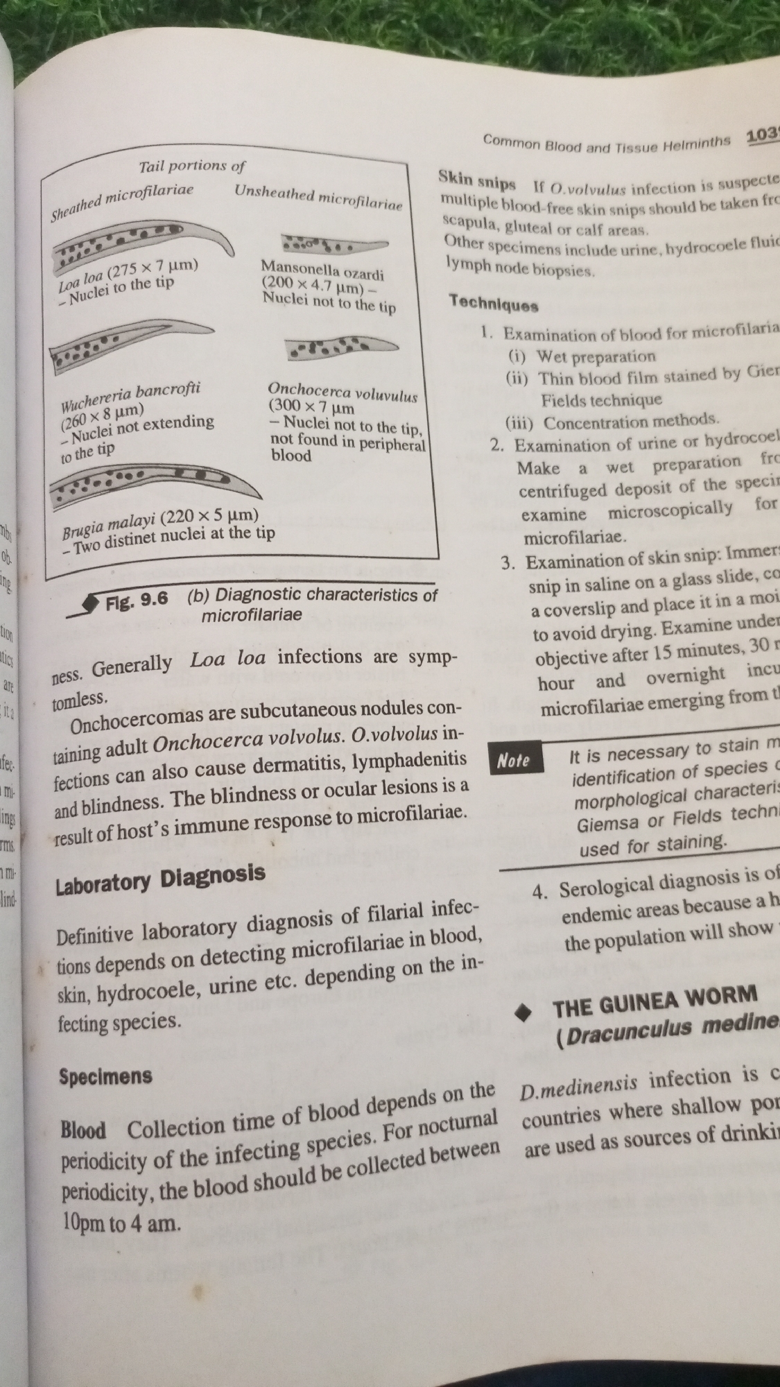
The image is a page from a medical textbook, likely a parasitology or tropical medicine section, detailing filarial infections. It contains text and illustrations. The text discusses "Common Blood and Tissue Helminths" and lists techniques for diagnosis. It describes "Skin snips" for suspected *O. volvulus* infections, mentioning that multiple blood-free skin snips should be taken from the scapula, gluteal, or calf areas. Other specimens mentioned include urine, hydrocoele fluid, and lymph node biopsies. Diagnostic techniques are numbered: 1. **Examination of blood for microfilariae:** Includes wet preparation, thin blood film stained by Giemsa or Fields technique, and concentration methods. 2. **Examination of urine or hydrocoele fluid:** Involves making a wet preparation from a centrifuged deposit and examining it microscopically for microfilariae. 3. **Examination of skin snip:** Describes immersing a skin snip in saline on a glass slide, covering it with a coverslip, and placing it in a moist chamber to avoid drying. Examination is to be done after 15 minutes, 30 minutes, 1 hour, and overnight to observe microfilariae emerging. A note specifies that staining with Giemsa or Fields technique is necessary for identifying species based on morphological characteristics. 4. **Serological diagnosis:** Mentioned for endemic areas where a high proportion of the population will show positive results. The image also includes a figure (Fig. 9.6) illustrating "Diagnostic characteristics of microfilariae." This figure shows diagrams of tail portions of different microfilariae, including: * **Loa loa:** Sheathed, with nuclei extending to the tip. * **Mansonella ozzardi:** Unsheathed, with nuclei not to the tip. * **Wuchereria bancrofti:** Sheathed, with nuclei not extending to the tip. * **Onchocerca volvulus:** Unsheathed, with nuclei not to the tip, not found in peripheral blood. * **Brugia malayi:** Sheathed, with two distinct nuclei at the tip. The text also describes *Loa loa* infections as generally asymptomatic. It mentions "Onchocercomas" as subcutaneous nodules containing adult *Onchocerca volvolus*, and *O. volvolus* infections can cause dermatitis, lymphadenitis, and blindness due to the host's immune response. Under "Laboratory Diagnosis," it states that definitive diagnosis depends on detecting microfilariae in blood, skin, hydrocoele, or urine, depending on the infecting species. Under "Specimens," it discusses "Blood Collection" and notes that the collection time depends on the nocturnal periodicity of the infecting species. For nocturnal periodicity, blood should be collected between 10 pm and 4 am. The text also has a section titled "THE GUINEA WORM (Dracunculus medinensis)" which begins to describe *D. medinensis* infection in
Symbol
D3DE7
Volume
6,956
Creator
+$0.01
Revenue
+$0.02
TVL
$0.93
Loading AttnAds…
2

Rev Bot 🤖💰
Injected revenue 2mo ago
“Revenue bonus for the last stake.”
+$0.03 USD