Stake attention in this memory
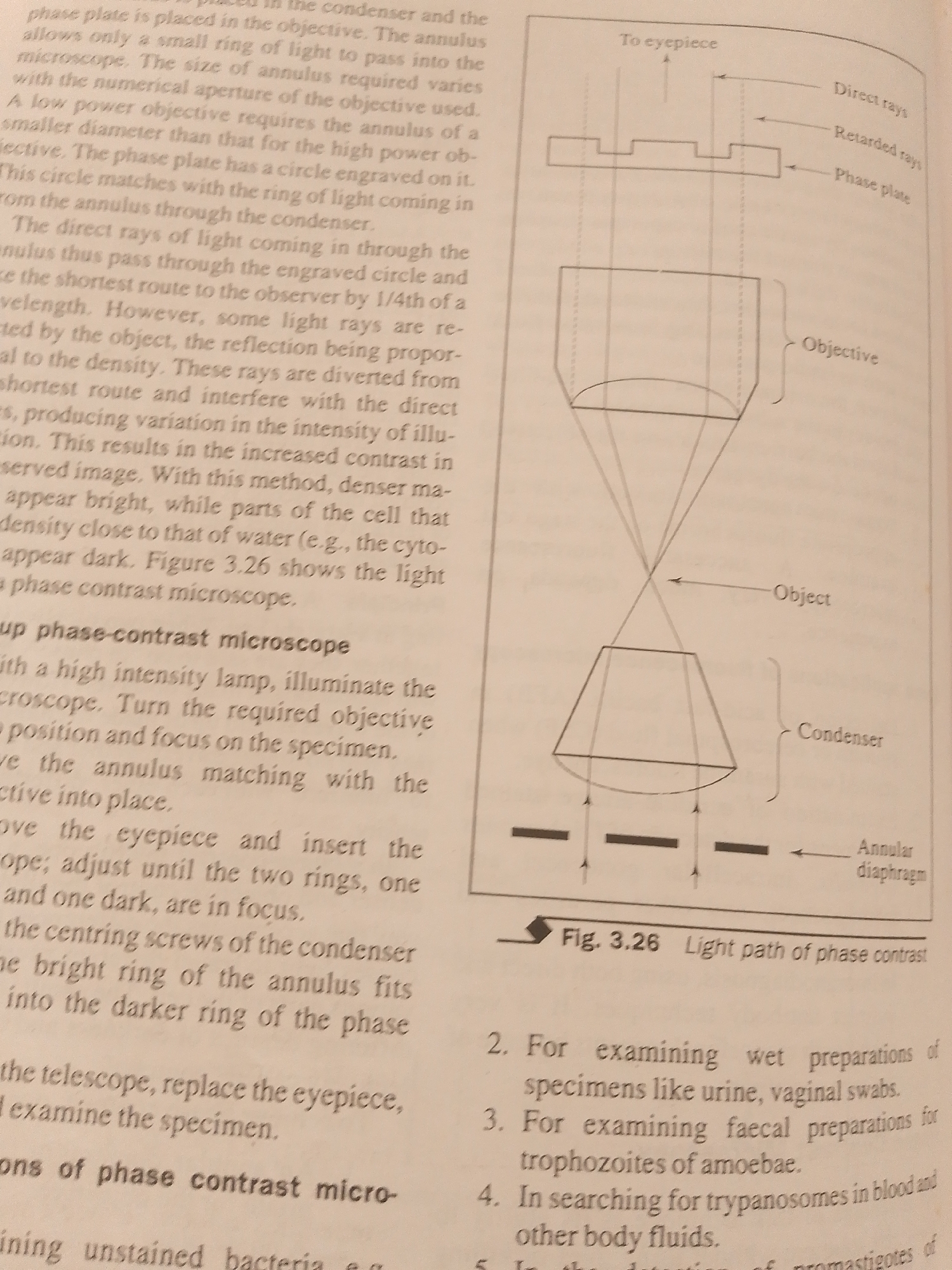
The image displays a page from a textbook or scientific document, focusing on the principles and operation of a phase-contrast microscope. The primary subjects are diagrams illustrating the light path through a phase-contrast microscope and accompanying text explaining its function and usage. The diagram on the right shows a schematic representation of light rays interacting with different components of the microscope, including the condenser, object, objective lens, and eyepiece. Labels like "Direct rays," "Retarded rays," "Phase plate," "Objective," "Object," "Condenser," and "Annular diaphragm" provide specific details about the optical path. The text on the left elaborates on these concepts, explaining how the phase plate and annulus work together to enhance contrast in unstained specimens. It details the process of setting up a phase-contrast microscope, including illuminating the specimen, adjusting the objective and annulus, and focusing. The text also lists applications of phase-contrast microscopy, such as examining wet preparations of urine and vaginal swabs, faecal preparations for amoebae, and searching for trypanosomes in blood and other body fluids. The scene is a static representation of scientific information, devoid of any indication of people, specific time of day, weather, or emotional context. The location context provided (Jalingo, Nigeria) is not visually represented in the image itself, which appears to be a generic educational illustration. The only visible text includes the diagram labels and the descriptive paragraphs, along with a figure caption: "Fig. 3.26 Light path of phase contrast."
No transactions found