
Stake attention in this memory
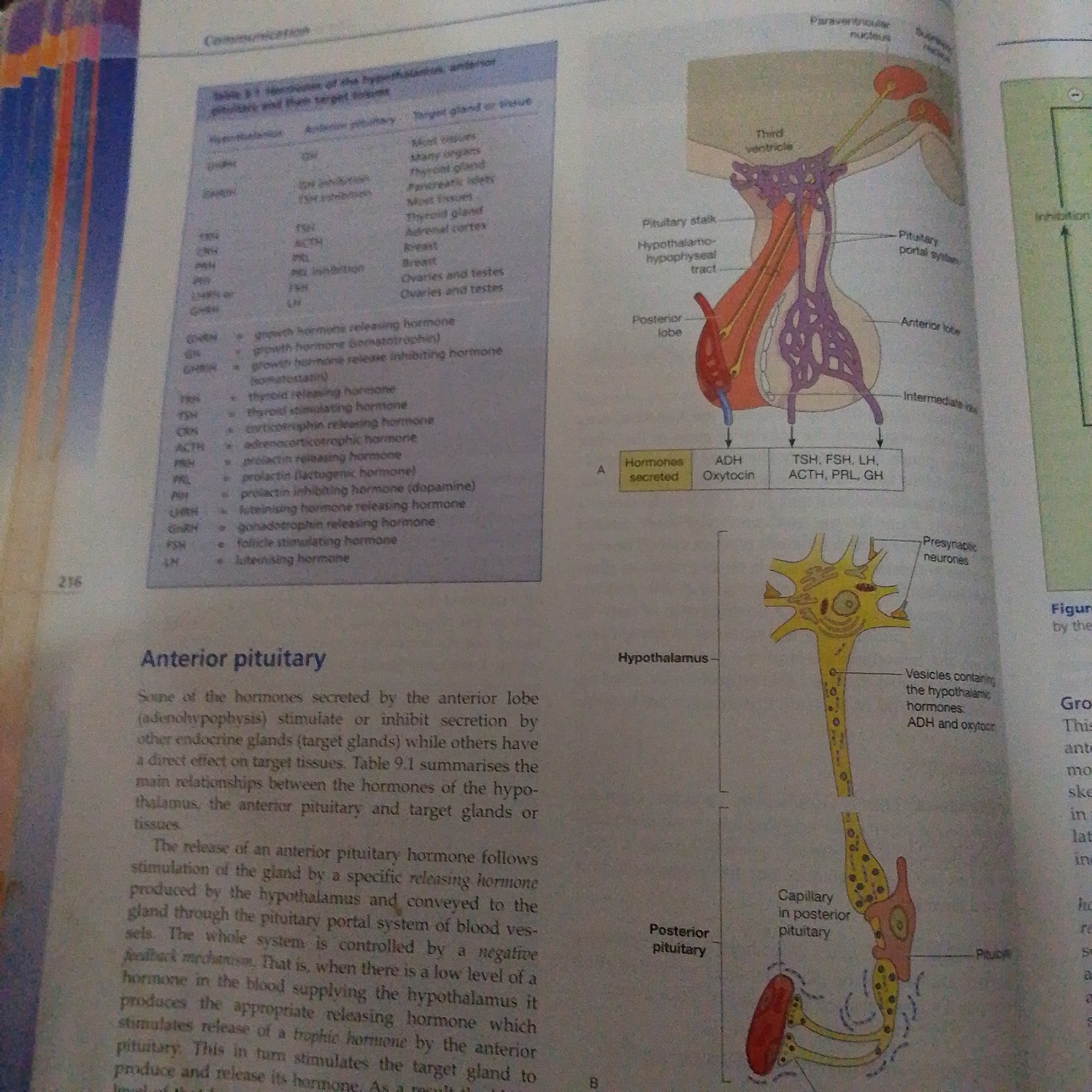
The image displays two diagrams illustrating the anatomy and hormonal regulation of the pituitary gland and its connection to the hypothalamus. The first diagram, on the upper right, shows a sagittal view of the hypothalamus and pituitary gland, highlighting the third ventricle, paraventricular nucleus, and supraoptic nucleus within the hypothalamus, as well as the pituitary stalk, posterior lobe, anterior lobe, and intermediate lobe of the pituitary. The second diagram, below the first, focuses on the posterior pituitary, depicting a neuron with its axon terminals in the posterior pituitary, showing vesicles containing ADH and oxytocin, and a capillary network. To the left of the diagrams is a table, labeled "Table 9.1 Hormones of the hypothalamus, anterior pituitary and their target tissues," which lists various hypothalamic and anterior pituitary hormones, their abbreviations, and their target glands or tissues. The text below the table discusses the anterior pituitary's role in stimulating or inhibiting other endocrine glands and the mechanism of hormone release, involving releasing hormones from the hypothalamus and negative feedback. The overall scene appears to be from a textbook or educational material, indicated by the presence of diagrams, tables, and explanatory text. There are no people or specific environmental cues to suggest a particular time of day, weather, or explicit location beyond what is implied by the scientific subject matter. The image is educational in nature, focused on explaining complex physiological processes. Visible text includes labels for anatomical structures, hormone names, and descriptive paragraphs.
No transactions found