
Sign in
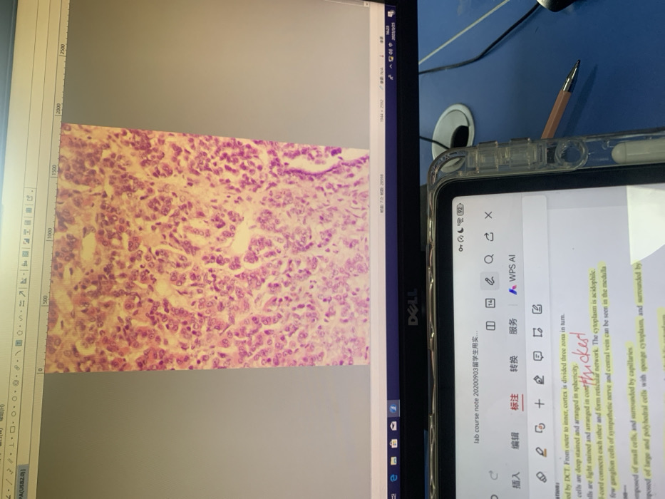
An image of a histological sample is displayed on a computer monitor. The tissue section is stained purple and pink, revealing cellular details. Next to the monitor, a tablet displays a document. The text on the tablet discusses the different zones of the cortex, noting how they are stained and arranged. One section is marked as "thickest". The cytoplasm of the cells is described as acidophilic, and ganglion cells and central veins are mentioned. The tissue consists of small cells surrounded by capillaries.
Emma🇨🇳💕

Dec 8, 2025, 1:24 AM

Shenyang City, China

Stake attention in this memory

microscopic image
histology
medical education
cell biology
cellular structure

An image of a histological sample is displayed on a computer monitor. The tissue section is stained purple and pink, revealing cellular details. Next to the monitor, a tablet displays a document. The text on the tablet discusses the different zones of the cortex, noting how they are stained and arranged. One section is marked as "thickest". The cytoplasm of the cells is described as acidophilic, and ganglion cells and central veins are mentioned. The tissue consists of small cells surrounded by capillaries.

Loading AttnAds…

transactions
revenues
stakers
Earliest
Latest
Highest stake

No transactions found
More from this user

More from Shenyang City

Emma🇨🇳💕

Dec 8, 2025, 1:24 AM

Shenyang City, China

Licensed under Creative Commons Attribution 4.0 International License. You are free to share and adapt this content with proper attribution.