Stake attention in this memory
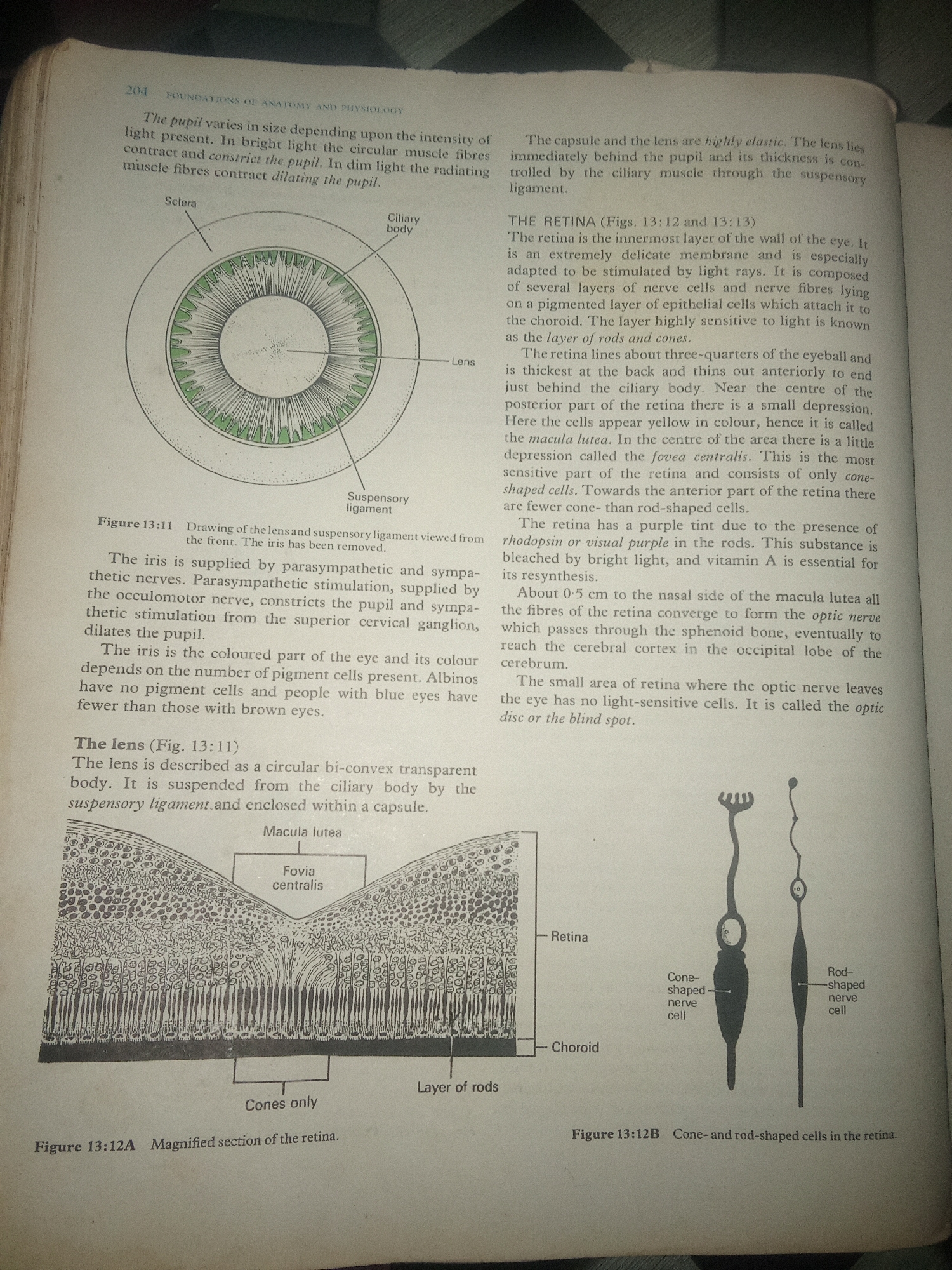
The image is a page from a textbook. It contains anatomical diagrams of the human eye and accompanying text. There are three illustrations: 1. **Figure 13:11** shows a cross-section of the iris, ciliary body, suspensory ligament, and lens, viewed from the front. 2. **Figure 13:12A** depicts a magnified cross-section of the retina, showing layers including the macula lutea, fovea centralis, and the layer of rods and cones. 3. **Figure 13:12B** illustrates cone-shaped and rod-shaped nerve cells found in the retina. The text discusses the iris, lens, retina, and its light-sensitive cells (rods and cones). There is no information or context provided that relates to Jalingo, Nigeria, or any urban environment. The image is solely an educational illustration.
Loading AttnAds…
No transactions found