
Sign in
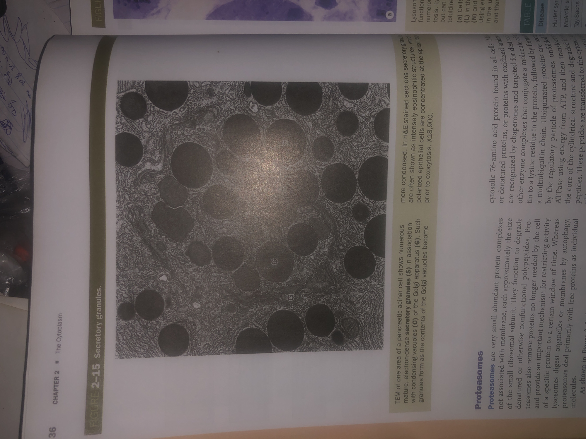
This image displays a textbook page. The top left corner shows the number "36" and the text "CHAPTER 2 The Cytoplasm". Below this, a section is titled "FIGURE 2-15 Secretory granules." This section includes a grayscale electron micrograph of numerous circular structures, identified as secretory granules, within a cell. The caption below the micrograph describes these granules as mature, electron-dense secretory granules (S) associated with condensing vacuoles (C) of the Golgi apparatus (G) in a pancreatic acinar cell. The text also notes that these granules become more condensed and are often shown as intensely eosinophilic structures in H&E-stained sections prior to exocytosis.

Below the micrograph, there is a section titled "Proteasomes." This text describes proteasomes as small, abundant protein complexes involved in degrading denatured or nonfunctional polypeptides. They are also mentioned as playing a role in autophagic breakdown of organelles and membranes.

The right side of the page has a section labeled "FIGURE" with a partial image of stained cells and accompanying text that is largely illegible. There is also a table with the heading "TABLE" and some text related to diseases.

The image provided does not contain any information or visual elements that would allow for a description of the city of Wukari, Nigeria, or any scene located there.
Bigstev

Jun 12, 2026, 1:42 AM

Wukari, Nigeria

Moment

Stake attention in this memory

scientific
informative
educational
detailed
technical

This image displays a textbook page. The top left corner shows the number "36" and the text "CHAPTER 2 The Cytoplasm". Below this, a section is titled "FIGURE 2-15 Secretory granules." This section includes a grayscale electron micrograph of numerous circular structures, identified as secretory granules, within a cell. The caption below the micrograph describes these granules as mature, electron-dense secretory granules (S) associated with condensing vacuoles (C) of the Golgi apparatus (G) in a pancreatic acinar cell. The text also notes that these granules become more condensed and are often shown as intensely eosinophilic structures in H&E-stained sections prior to exocytosis. Below the micrograph, there is a section titled "Proteasomes." This text describes proteasomes as small, abundant protein complexes involved in degrading denatured or nonfunctional polypeptides. They are also mentioned as playing a role in autophagic breakdown of organelles and membranes. The right side of the page has a section labeled "FIGURE" with a partial image of stained cells and accompanying text that is largely illegible. There is also a table with the heading "TABLE" and some text related to diseases. The image provided does not contain any information or visual elements that would allow for a description of the city of Wukari, Nigeria, or any scene located there.

Symbol

E1FBF

Volume

11,285

Creator

+$0.03

Revenue

+$0.04

TVL

$2.80

1D
1W
1M
All

Loading AttnAds…

transactions
revenues
stakers
Earliest
Latest
Highest stake

1

ZaddyTanko

Staked 26d ago

$2.79 USD

+$0.01 USD

+0.4%

2

Rev Bot 🤖💰

Injected revenue 26d ago

Revenue bonus for the last stake.

+$0.04 USD

More from this user

More from Wukari

Bigstev

Jun 12, 2026, 1:42 AM

Wukari, Nigeria

Licensed under Creative Commons Attribution 4.0 International License. You are free to share and adapt this content with proper attribution.