
Stake attention in this memory
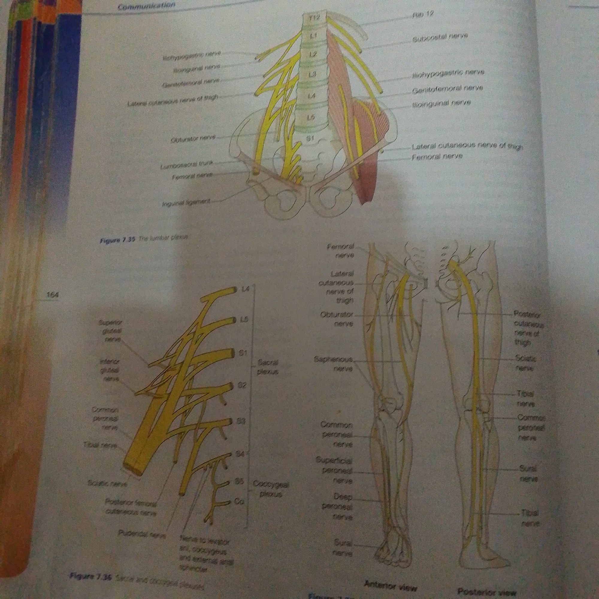
The image displays an anatomical illustration of the lumbar and sacral plexuses, along with their associated nerves, presented in a textbook or educational material. The primary subject is the detailed diagram of the nervous system in the pelvic and leg regions, rendered in yellow on a white background. There are no people or a discernible real-world scene depicted; the context is purely illustrative. The diagram is divided into several parts. One section shows the lumbar spine (labeled T12, L1-L5, S1) with nerves branching out towards the anterior and lateral aspects of the hip and thigh. Labels like "Iliohypogastric nerve," "Genitofemoral nerve," "Lateral cutaneous nerve of thigh," and "Femoral nerve" identify these structures. Another section illustrates the nerves of the lower leg and thigh from both an anterior and posterior view. Visible labels include "Sciatic nerve," "Tibial nerve," "Common peroneal nerve," and "Saphenous nerve." A smaller diagram shows the sacral plexus (labeled S1-S5, Co) and its branches. There is visible text in the image, including figure captions like "Figure 7.35 The lumbar plexus" and "Figure 7.36 Sacral and coccygeal plexus," as well as page numbers like "164." The overall presentation suggests an educational setting, likely a medical or anatomy textbook. No specific time of day, weather, or emotional cues are present. The location context provided (Jalingo, Nigeria) is not visually evident within the image itself.
Loading AttnAds…
No transactions found