
Stake attention in this memory
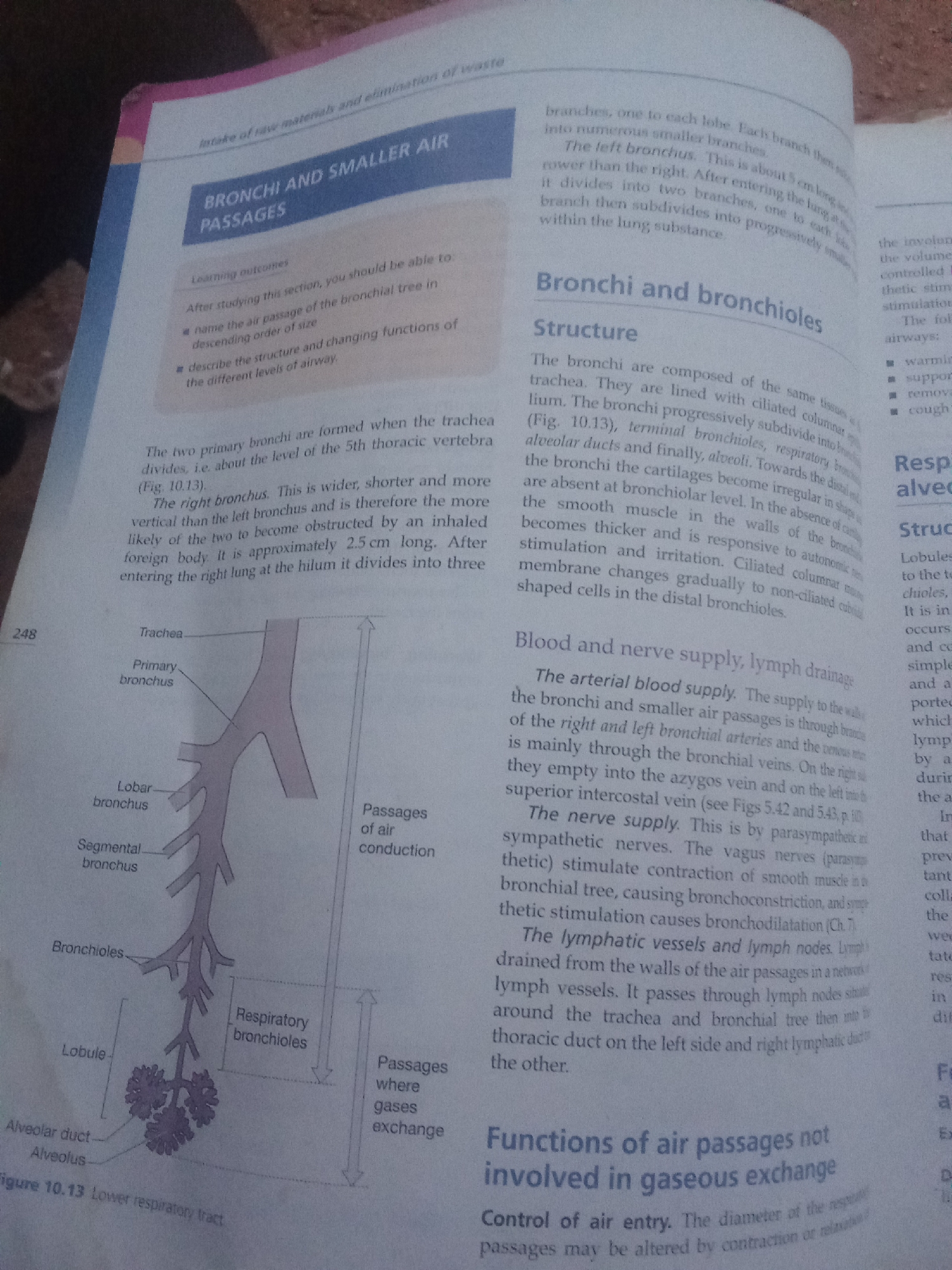
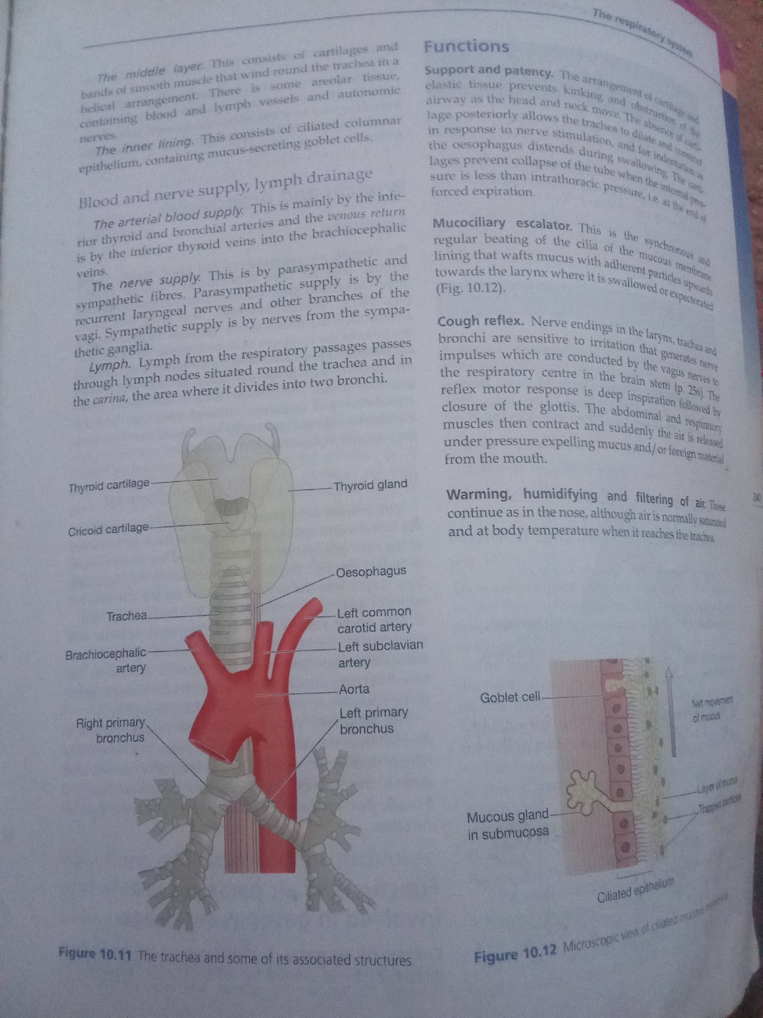
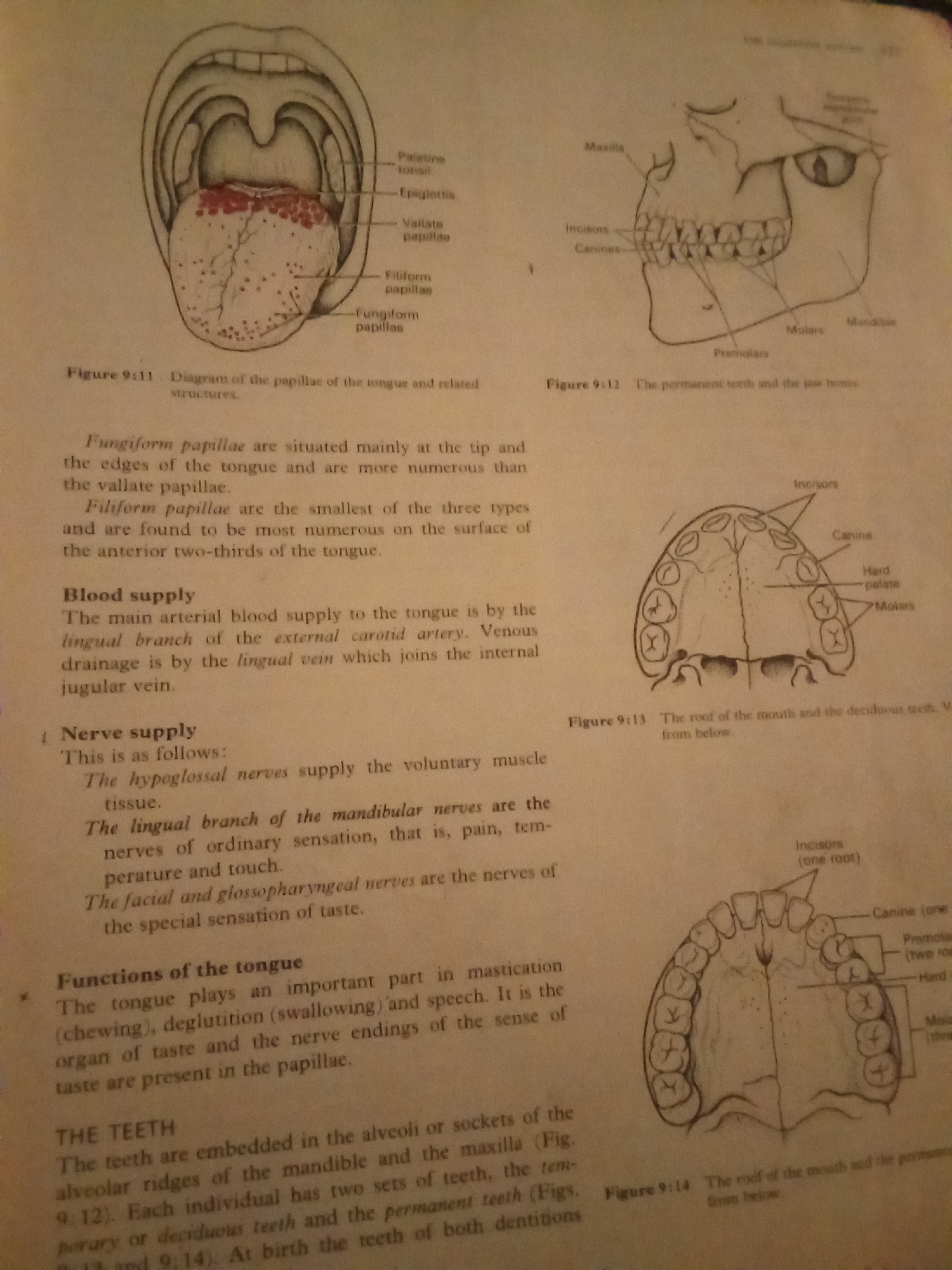
The image displays anatomical illustrations of the human mouth and teeth, accompanied by descriptive text. The first illustration, labeled Figure 9:11, shows a diagram of the tongue with various types of papillae, including palatine tonsil, epiglottis, vallate papillae, filiform papillae, and fungiform papillae. The second illustration, Figure 9:12, depicts a lateral view of the skull, highlighting the permanent teeth and jawbones, with labels for maxilla, mandible, incisors, canines, premolars, and molars. The text below these figures describes the location and characteristics of fungiform and filiform papillae, the blood supply to the tongue via the lingual artery and vein, and the nerve supply to the tongue by the hypoglossal, mandibular, facial, and glossopharyngeal nerves. It also mentions the functions of the tongue, such as mastication, deglutition, and speech, as well as its role in taste sensation. The third illustration, Figure 9:13, provides a superior view of the roof of the mouth and deciduous teeth, with labels for incisors, canines, hard palate, and molars. The final illustration, Figure 9:14, shows a superior view of the roof of the mouth and permanent teeth, again with labels for incisors, canines, premolars, hard palate, and molars. The accompanying text discusses the embedding of teeth in the alveoli of the mandible and maxilla, and the presence of two sets of teeth in individuals: deciduous and permanent. The image does not contain any information that would allow for its location in Jalingo, Nigeria, or any other geographical location. The content is purely anatomical and educational.
Loading AttnAds…
No transactions found