Sign in
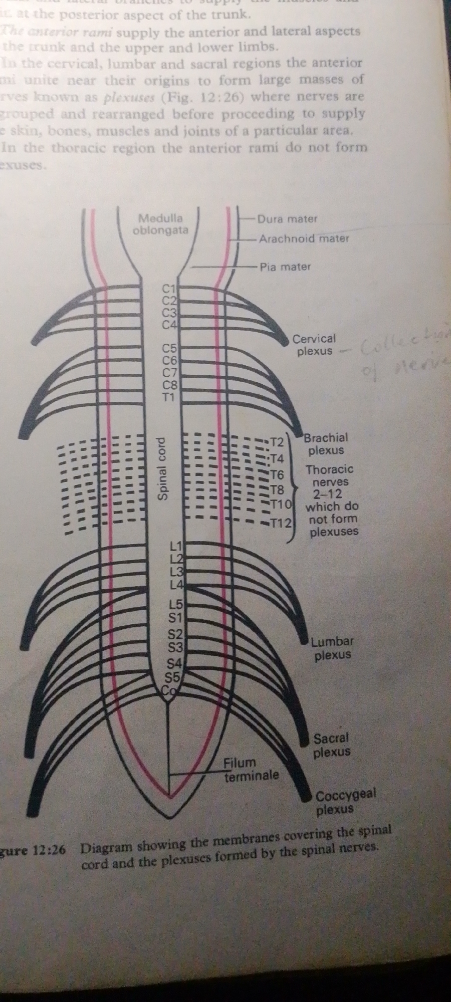
This media file is an image of a diagram from a textbook, illustrating the human spinal cord and the nervous plexuses formed by spinal nerves. The diagram shows the spinal cord extending downwards from the medulla oblongata, with the surrounding protective membranes (dura mater, arachnoid mater, and pia mater). Spinal nerves are depicted branching out from the cord at different vertebral levels (C1-C8, T1-T12, L1-L5, S1-S5, and Co). The diagram specifically highlights the formation of the cervical, brachial, lumbar, sacral, and coccygeal plexuses, which are networks of nerves that branch and rejoin to supply specific areas of the body. The thoracic nerves T2-T12 are noted as not forming plexuses. There is also a label for the "Filum terminale," a filament extending from the end of the spinal cord.

The text within the image provides context and explanations for the diagram. It describes how anterior rami of spinal nerves unite to form plexuses in the cervical, lumbar, and sacral regions, facilitating their rearrangement before supplying skin, bones, muscles, and joints. It also states that anterior rami in the thoracic region do not form plexuses. The caption at the bottom reads "Figure 12:26 Diagram showing the membranes covering the spinal cord and the plexuses formed by the spinal nerves." The visible text also includes handwritten annotations, such as "Cervical plexus - Collection of nerve".

The image is an educational illustration and does not depict any people, a specific scene, setting, or event in Jauro Yinu, Nigeria. It is a purely anatomical and physiological representation.
FM-2L6Lq2

Jun 30, 2026, 4:52 PM

Jauro Yinu, Nigeria

Moment

Stake attention in this memory

studious
analytical
focused
curious
academic

This media file is an image of a diagram from a textbook, illustrating the human spinal cord and the nervous plexuses formed by spinal nerves. The diagram shows the spinal cord extending downwards from the medulla oblongata, with the surrounding protective membranes (dura mater, arachnoid mater, and pia mater). Spinal nerves are depicted branching out from the cord at different vertebral levels (C1-C8, T1-T12, L1-L5, S1-S5, and Co). The diagram specifically highlights the formation of the cervical, brachial, lumbar, sacral, and coccygeal plexuses, which are networks of nerves that branch and rejoin to supply specific areas of the body. The thoracic nerves T2-T12 are noted as not forming plexuses. There is also a label for the "Filum terminale," a filament extending from the end of the spinal cord. The text within the image provides context and explanations for the diagram. It describes how anterior rami of spinal nerves unite to form plexuses in the cervical, lumbar, and sacral regions, facilitating their rearrangement before supplying skin, bones, muscles, and joints. It also states that anterior rami in the thoracic region do not form plexuses. The caption at the bottom reads "Figure 12:26 Diagram showing the membranes covering the spinal cord and the plexuses formed by the spinal nerves." The visible text also includes handwritten annotations, such as "Cervical plexus - Collection of nerve". The image is an educational illustration and does not depict any people, a specific scene, setting, or event in Jauro Yinu, Nigeria. It is a purely anatomical and physiological representation.

Symbol

E938F

Volume

9,373

Creator

+$0.02

Revenue

+$0.04

TVL

$1.97

1D
1W
1M
All

Loading AttnAds…

transactions
revenues
stakers
Earliest
Latest
Highest stake

1

FM-FSRgn1

Staked 16d ago

$1.95 USD

+$0.02 USD

+1.0%

2

Rev Bot 🤖💰

Injected revenue 16d ago

Revenue bonus for the last stake.

+$0.04 USD

More from this user

More from Jauro Yinu

FM-2L6Lq2

Jun 30, 2026, 4:52 PM

Jauro Yinu, Nigeria

Licensed under Creative Commons Attribution 4.0 International License. You are free to share and adapt this content with proper attribution.