
Sign in
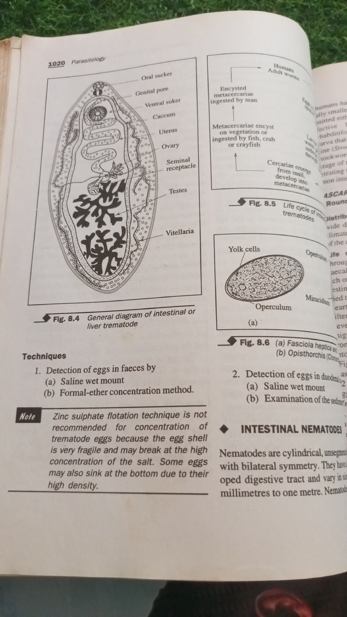
The image is a page from a biology textbook discussing parasitology. It features several diagrams and text.

On the left, Figure 8.4 is a detailed diagram of an intestinal or liver trematode, with various parts labeled such as "Oral sucker," "Ventral sucker," "Caccum," "Uterus," "Ovary," "Seminal receptacle," "Testes," and "Vitellaria." Below this diagram, the caption reads "Fig. 8.4 General diagram of intestinal or liver trematode."

To the right of the trematode diagram, there is a life cycle diagram labeled "Fig. 8.5 Life cycle of intestinal trematodes." This diagram illustrates stages including "Encycsted metacercariae ingested by man," "Metacercariae encyst on vegetation or ingested by fish, crab or crayfish," and "Cercariae emerge from snail, develop into metacercariae." It also shows "Adult worms" for humans.

Below the trematode diagram and life cycle, there is a section titled "Techniques." It lists "1. Detection of eggs in faeces by (a) Saline wet mount (b) Formal-ether concentration method."

A "Note" box provides information: "Zinc sulphate flotation technique is not recommended for concentration of trematode eggs because the egg shell is very fragile and may break at the high concentration of the salt. Some eggs may also sink at the bottom due to their high density."

Further to the right, Figure 8.6 shows an illustration of a trematode egg. The caption reads "Fig. 8.6 (a) Fasciola hepatica and (b) Opisthorchis (Clonorchis)."

Below Figure 8.6, there is a point numbered "2. Detection of eggs in duodenal fluid (a) Saline wet mount (b) Examination of the sediment."

At the bottom right, there is a section heading "INTESTINAL NEMATODES." The text below describes nematodes as "cylindrical, unsegmented with bilateral symmetry. They have a developed digestive tract and vary in size from millimetres to one metre."

The image is a scanned page, likely from a book or document, with the texture of paper visible. The background above and to the right of the page appears to be a blurry green surface, suggesting it was photographed on a table or surface covered with a green material, perhaps grass or a mat. The image is brightly lit, and some text is slightly blurred due to the angle of the photograph.

There is no information within the image that relates to the city of Jalingo, Nigeria.
FM-zwv5T2

Jun 28, 2026, 11:32 AM

Jalingo, Nigeria

Short Term

Stake attention in this memory

educational
scientific
informative
detailed
academic

The image is a page from a biology textbook discussing parasitology. It features several diagrams and text. On the left, Figure 8.4 is a detailed diagram of an intestinal or liver trematode, with various parts labeled such as "Oral sucker," "Ventral sucker," "Caccum," "Uterus," "Ovary," "Seminal receptacle," "Testes," and "Vitellaria." Below this diagram, the caption reads "Fig. 8.4 General diagram of intestinal or liver trematode." To the right of the trematode diagram, there is a life cycle diagram labeled "Fig. 8.5 Life cycle of intestinal trematodes." This diagram illustrates stages including "Encycsted metacercariae ingested by man," "Metacercariae encyst on vegetation or ingested by fish, crab or crayfish," and "Cercariae emerge from snail, develop into metacercariae." It also shows "Adult worms" for humans. Below the trematode diagram and life cycle, there is a section titled "Techniques." It lists "1. Detection of eggs in faeces by (a) Saline wet mount (b) Formal-ether concentration method." A "Note" box provides information: "Zinc sulphate flotation technique is not recommended for concentration of trematode eggs because the egg shell is very fragile and may break at the high concentration of the salt. Some eggs may also sink at the bottom due to their high density." Further to the right, Figure 8.6 shows an illustration of a trematode egg. The caption reads "Fig. 8.6 (a) Fasciola hepatica and (b) Opisthorchis (Clonorchis)." Below Figure 8.6, there is a point numbered "2. Detection of eggs in duodenal fluid (a) Saline wet mount (b) Examination of the sediment." At the bottom right, there is a section heading "INTESTINAL NEMATODES." The text below describes nematodes as "cylindrical, unsegmented with bilateral symmetry. They have a developed digestive tract and vary in size from millimetres to one metre." The image is a scanned page, likely from a book or document, with the texture of paper visible. The background above and to the right of the page appears to be a blurry green surface, suggesting it was photographed on a table or surface covered with a green material, perhaps grass or a mat. The image is brightly lit, and some text is slightly blurred due to the angle of the photograph. There is no information within the image that relates to the city of Jalingo, Nigeria.

Symbol

EA1D6

Volume

11,550

Creator

+$0.03

Revenue

+$0.04

TVL

$2.55

1D
1W
1M
All

Loading AttnAds…

transactions
revenues
stakers
Earliest
Latest
Highest stake

1

FM-FzB8r2

Staked 2mo ago

$2.53 USD

+$0.02 USD

+0.6%

2

Rev Bot 🤖💰

Injected revenue 2mo ago

Revenue bonus for the last stake.

+$0.05 USD

More from this user

More from Jalingo

FM-zwv5T2

Jun 28, 2026, 11:32 AM

Jalingo, Nigeria

Licensed under Creative Commons Attribution 4.0 International License. You are free to share and adapt this content with proper attribution.