
Stake attention in this memory
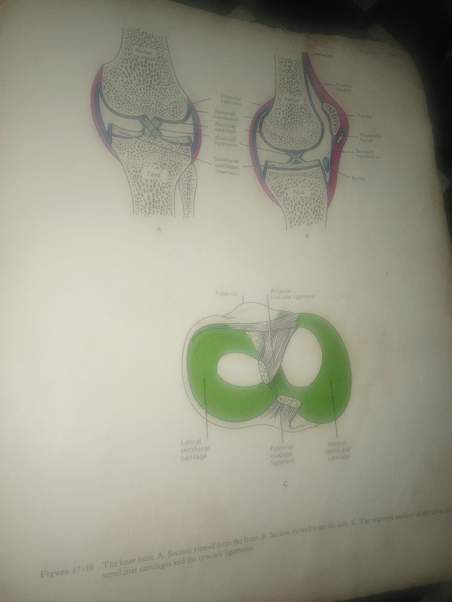
The image displays three diagrams illustrating the anatomy of the human knee joint. Diagram A shows a frontal section of the knee, labeling components such as the femur, tibia, capsular ligament, synovial membrane, articular cartilage, cruciate ligaments, and semilunar cartilages. Diagram B presents a side section of the knee, indicating the femur, patella, patellar tendon, prepatellar bursa, synovial membrane, and bursa. Diagram C offers a superior view of the tibia, showing the lateral and medial semilunar cartilages, as well as the anterior and posterior cruciate ligaments. The diagrams are accompanied by the caption "Figure 17:10 The knee joint. A. Section viewed from the front. B. Section viewed from the side. C. The superior surface of the tibia showing semilunar cartilages and the cruciate ligaments." There is no information in the image that relates to the city of Jalingo, Nigeria, or any specific location. The image is purely an anatomical illustration.
Loading AttnAds…
No transactions found