Stake attention in this memory
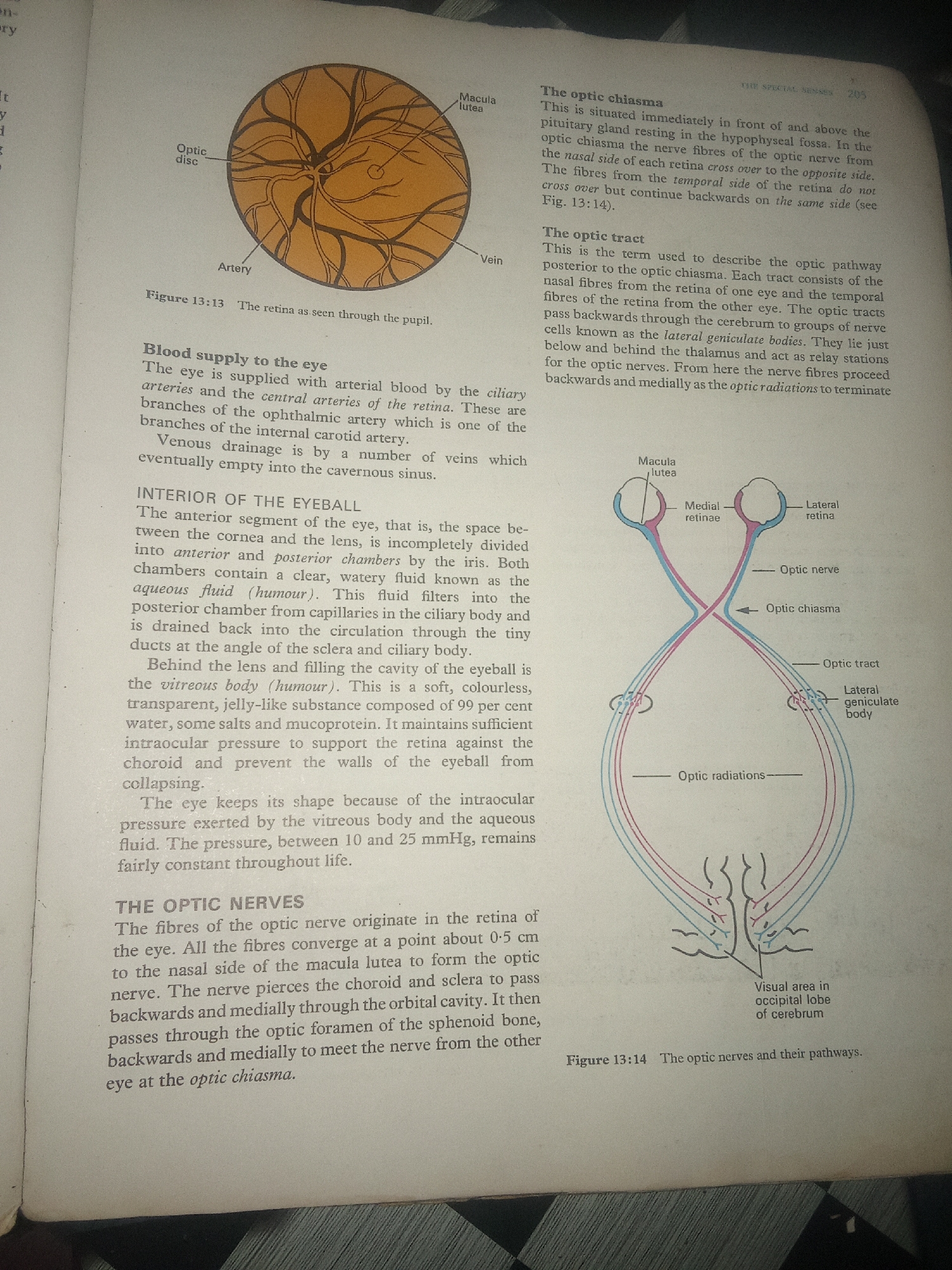
This image is a scanned page from a textbook, likely detailing the anatomy and physiology of the human eye. The primary subject is the eye and its neural pathways. The page contains two diagrams. The first, labeled Figure 13:13, shows a cross-section of the retina as seen through the pupil, illustrating the optic disc, macula lutea, artery, and vein. The second, Figure 13:14, depicts the optic nerves and their pathways, showing the optic chiasma, optic tract, lateral geniculate body, and the visual area in the occipital lobe of the cerebrum. The diagrams use color to differentiate nerve fibers. Accompanying the diagrams are several blocks of text that explain various aspects of the eye, including: - **Blood supply to the eye**: Discussing arterial and venous drainage. - **Interior of the eyeball**: Describing the anterior and posterior chambers, aqueous humor, and vitreous body. - **The optic chiasma**: Explaining the crossing of nerve fibers. - **The optic tract**: Detailing the pathway from the optic chiasma to the lateral geniculate bodies. - **The optic nerves**: Describing the origin and path of the optic nerve fibers. There is also some text visible on the left margin of the page, which appears to be from a previous section of the textbook. The text on the right margin includes a page number "205" and a heading "THE SPECIAL SENSES". The overall scene is that of an educational document, presenting scientific information through text and illustrations. There are no people, animals, or a specific natural setting depicted, other than the implied biological subject matter. The lighting appears to be neutral, consistent with a scanned document under artificial light. The image is taken from a slightly angled perspective, showing the page within a book.
Loading AttnAds…
No transactions found