
Stake attention in this memory
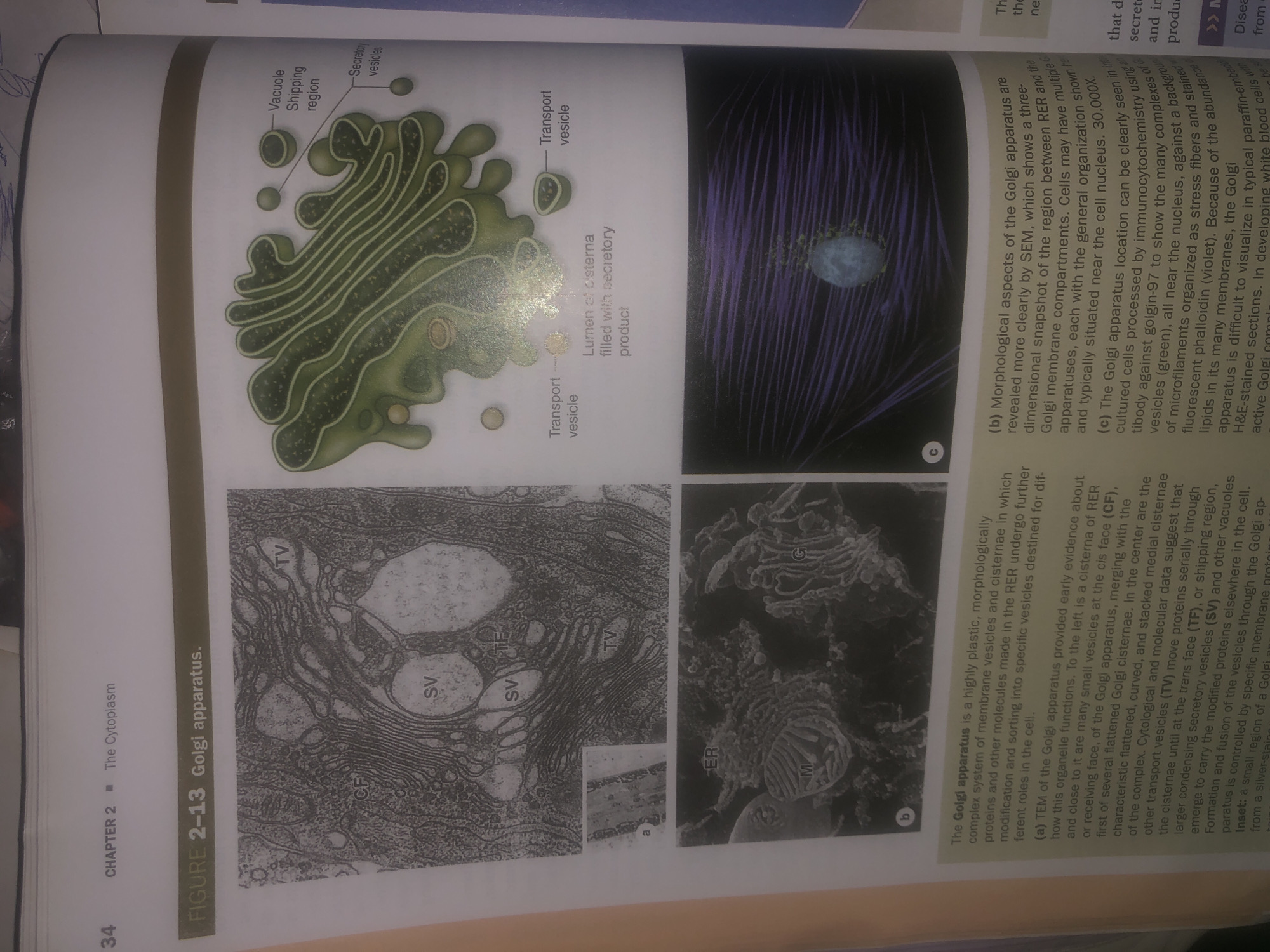
The image displays Figure 2-13, illustrating the Golgi apparatus. It includes three panels: Panel (a) is a transmission electron micrograph (TEM) showing the ultrastructure of the Golgi apparatus, with labels for cis face (CF), cisternae (TV), and secretory vesicles (SV). Panel (b) is a diagrammatic representation of the Golgi apparatus, highlighting its stacked cisternae, transport vesicles, vacuole shipping region, and secretory vesicles. Panel (c) is a scanning electron micrograph (SEM) of the Golgi apparatus within a cell, showing its three-dimensional morphology in relation to other cellular components, including the endoplasmic reticulum (ER) and mitochondria (M). The accompanying text provides a detailed description of the Golgi apparatus, its function in modifying, sorting, and packaging proteins and lipids, and the evidence from TEM and SEM studies. There is no information in the image or text that relates to Wukari, Nigeria.
Symbol
F09AF
Volume
10,767
Creator
+$0.03
Revenue
+$0.05
TVL
$2.51
Loading AttnAds…

2

Rev Bot 🤖💰
Injected revenue 26d ago
“Revenue bonus for the last stake.”
+$0.05 USD