Sign in
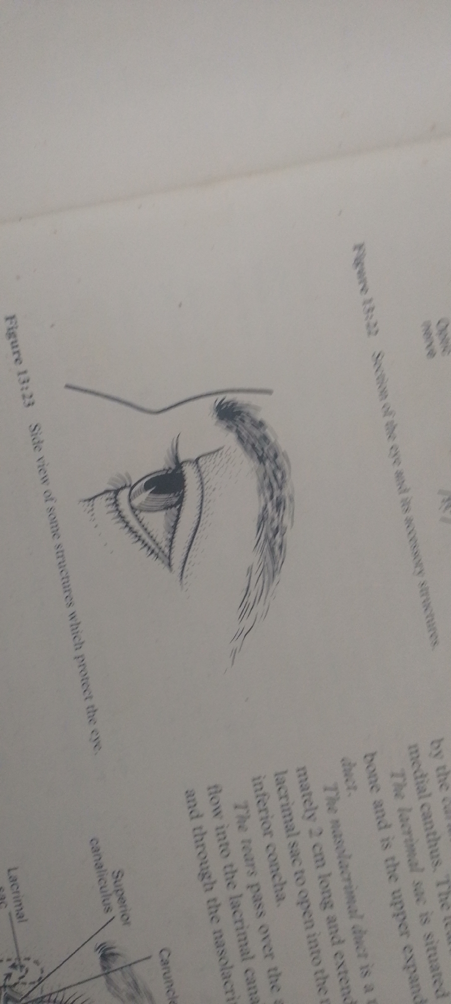
This media file shows an illustration of the human eye and its surrounding structures, likely from a textbook or anatomical reference. The drawing depicts a side view of an eye, highlighting the eyelid, eyelashes, iris, and pupil. Lines and labels indicate various anatomical features such as the superior canaliculus and lacrimal sac.

The image also includes text that appears to be captions and descriptive labels. One caption reads "Figure 13:23 Side view of some structures which protect the eye." Another caption, "Figure 13:22 Section of the eye and its accessory structures," is also visible. There are labels for "Optic nerve," "Superior canaliculus," "Lacrimal sac," and "Caruncle." The text also describes the path of tears from the eye, mentioning the lacrimal sac, nasolacrimal duct, and inferior concha.

The overall presentation suggests an educational context, aiming to inform the viewer about the anatomy and function of the eye. The drawing style is detailed and illustrative, typical of scientific diagrams. There are no people or specific environmental settings depicted, only the anatomical illustration and accompanying text. The time of day, weather, and location are not discernible from the image.
FM-2L6Lq2

Jun 30, 2026, 5:22 PM

Jauro Yinu, Nigeria

Moment

Stake attention in this memory

curious
analytical
focused
informative
educational

This media file shows an illustration of the human eye and its surrounding structures, likely from a textbook or anatomical reference. The drawing depicts a side view of an eye, highlighting the eyelid, eyelashes, iris, and pupil. Lines and labels indicate various anatomical features such as the superior canaliculus and lacrimal sac. The image also includes text that appears to be captions and descriptive labels. One caption reads "Figure 13:23 Side view of some structures which protect the eye." Another caption, "Figure 13:22 Section of the eye and its accessory structures," is also visible. There are labels for "Optic nerve," "Superior canaliculus," "Lacrimal sac," and "Caruncle." The text also describes the path of tears from the eye, mentioning the lacrimal sac, nasolacrimal duct, and inferior concha. The overall presentation suggests an educational context, aiming to inform the viewer about the anatomy and function of the eye. The drawing style is detailed and illustrative, typical of scientific diagrams. There are no people or specific environmental settings depicted, only the anatomical illustration and accompanying text. The time of day, weather, and location are not discernible from the image.

Symbol

F1B79

Volume

3,081

Creator

+$0.00

Revenue

+$0.01

TVL

$0.26

1D
1W
1M
All

Loading AttnAds…

transactions
revenues
stakers
Earliest
Latest
Highest stake

1

FM-FSRgn1

Staked 16d ago

$0.26 USD

+$0.00 USD

+1%

2

Rev Bot 🤖💰

Injected revenue 16d ago

Revenue bonus for the last stake.

+$0.01 USD

More from this user

More from Jauro Yinu

FM-2L6Lq2

Jun 30, 2026, 5:22 PM

Jauro Yinu, Nigeria

Licensed under Creative Commons Attribution 4.0 International License. You are free to share and adapt this content with proper attribution.