Sign in
The image shows a page from a medical textbook or exam paper, likely related to radiology. The primary visual element is a CT scan of a human head, displaying a cross-section with a prominent abnormality in the right parietal lobe. The CT scan is viewed from above.

The text on the page describes a clinical scenario involving a 24-year-old motorcyclist who has been in a road traffic accident and presents with confusion, localization of painful stimuli, and eye opening to painful stimuli. This scenario is followed by five questions asking to describe the CT, provide a diagnosis, identify the involved vessel, state the patient's Glasgow Coma Scale (GCS), and outline the management.

Below the questions are handwritten answers. The GCS calculation is written as "2 + 4 + 5 = 11" and "2 V + M", suggesting an attempt to score verbal and motor responses. The numbered answers indicate that the CT shows a bright, biconvex density in the right parietal lobe, identifying it as an Extradural Hematoma, and the Middle meningeal artery as the commonly involved vessel. The GCS is broken down into components: Confused (4), localize painful stimuli (5), and eye opening with pain (2).

The management options are listed as: a) Follow ATLS guidelines (A,B,C,D,E), b) Craniotomy and evacuation of hematoma, and c) Manage the patient in intensive care unit postoperatively. The page is numbered '59' at the bottom. The overall context is educational, focusing on the diagnosis and management of traumatic brain injuries, specifically an extradural hematoma. The lighting suggests the photo was taken indoors, likely in a study or examination room.
FM-xxDv52

Jun 17, 2026

Mansehra, Pakistan

BLOCKED

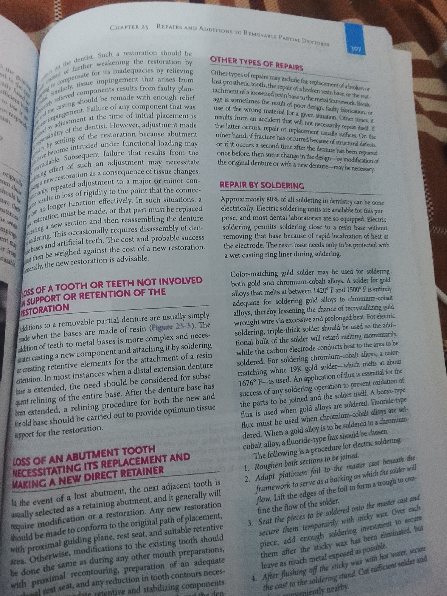
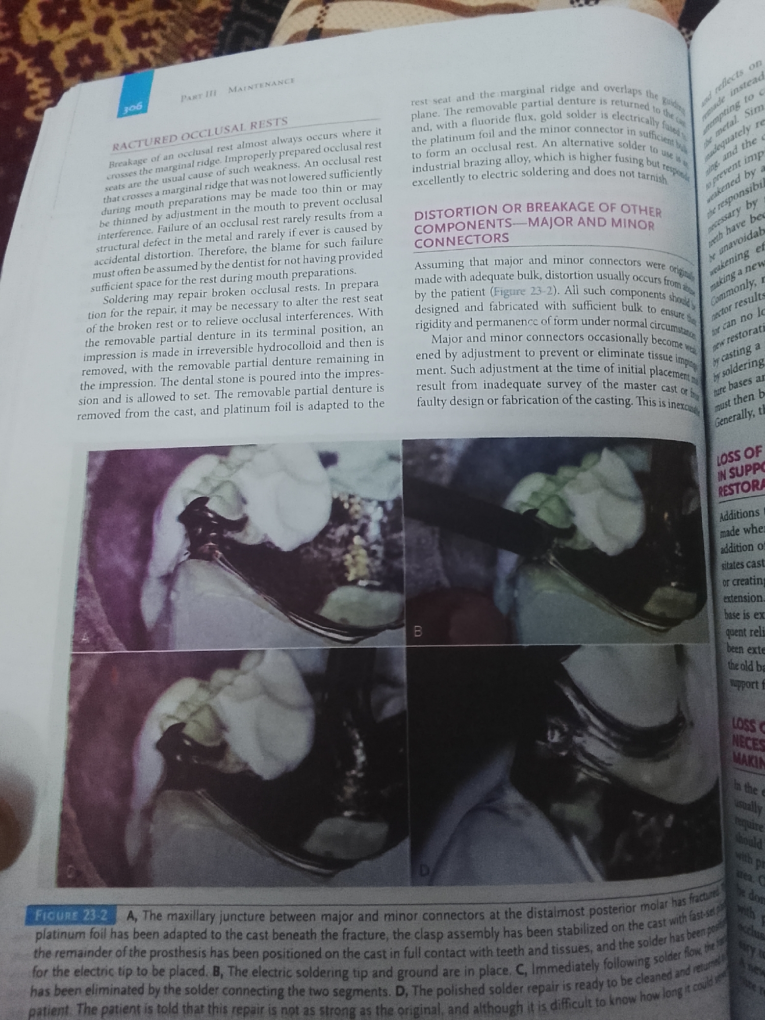
Stake attention in this memory

concerned
clinical
anxious
medical
serious

The image shows a page from a medical textbook or exam paper, likely related to radiology. The primary visual element is a CT scan of a human head, displaying a cross-section with a prominent abnormality in the right parietal lobe. The CT scan is viewed from above. The text on the page describes a clinical scenario involving a 24-year-old motorcyclist who has been in a road traffic accident and presents with confusion, localization of painful stimuli, and eye opening to painful stimuli. This scenario is followed by five questions asking to describe the CT, provide a diagnosis, identify the involved vessel, state the patient's Glasgow Coma Scale (GCS), and outline the management. Below the questions are handwritten answers. The GCS calculation is written as "2 + 4 + 5 = 11" and "2 V + M", suggesting an attempt to score verbal and motor responses. The numbered answers indicate that the CT shows a bright, biconvex density in the right parietal lobe, identifying it as an Extradural Hematoma, and the Middle meningeal artery as the commonly involved vessel. The GCS is broken down into components: Confused (4), localize painful stimuli (5), and eye opening with pain (2). The management options are listed as: a) Follow ATLS guidelines (A,B,C,D,E), b) Craniotomy and evacuation of hematoma, and c) Manage the patient in intensive care unit postoperatively. The page is numbered '59' at the bottom. The overall context is educational, focusing on the diagnosis and management of traumatic brain injuries, specifically an extradural hematoma. The lighting suggests the photo was taken indoors, likely in a study or examination room.

Symbol

F5DF3

Volume

11,550

Creator

+$0.20

Revenue

+$0.37

TVL

$15.64

1D
1W
1M
All
transactions
revenues
stakers
Earliest
Latest
Highest stake

1

FM-35bb12

Staked 10d ago

$15.42 USD

+$0.22 USD

+1%

2

Rev Bot 🤖💰

Injected revenue 10d ago

Revenue bonus for the last stake.

+$0.42 USD

More from this user

More from Mansehra

FM-xxDv52

Jun 17, 2026

Mansehra, Pakistan

Licensed under Creative Commons Attribution 4.0 International License. You are free to share and adapt this content with proper attribution.