
Sign in
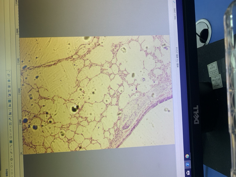
The image displays a microscopic view of lung tissue. Alveoli, the tiny air sacs responsible for gas exchange, are clearly visible as small, rounded structures. Thin walls of alveolar septa separate them. There is a thin layer of cells lining the outermost region. Overall, the lung structure appears somewhat disorganized or congested, possibly indicating a pathological condition. The image is captured on a computer screen displaying image editing software and a Dell monitor is visible on the right.
Emma🇨🇳💕

Nov 24, 2025, 2:04 AM

Shenyang City, China

Stake attention in this memory

microscopy
histology
lung tissue
alveolar sacs
stained cells

The image displays a microscopic view of lung tissue. Alveoli, the tiny air sacs responsible for gas exchange, are clearly visible as small, rounded structures. Thin walls of alveolar septa separate them. There is a thin layer of cells lining the outermost region. Overall, the lung structure appears somewhat disorganized or congested, possibly indicating a pathological condition. The image is captured on a computer screen displaying image editing software and a Dell monitor is visible on the right.

Loading AttnAds…

transactions
revenues
stakers
Earliest
Latest
Highest stake

No transactions found
More from this user

More from Shenyang City

Emma🇨🇳💕

Nov 24, 2025, 2:04 AM

Shenyang City, China

Licensed under Creative Commons Attribution 4.0 International License. You are free to share and adapt this content with proper attribution.