
Stake attention in this memory
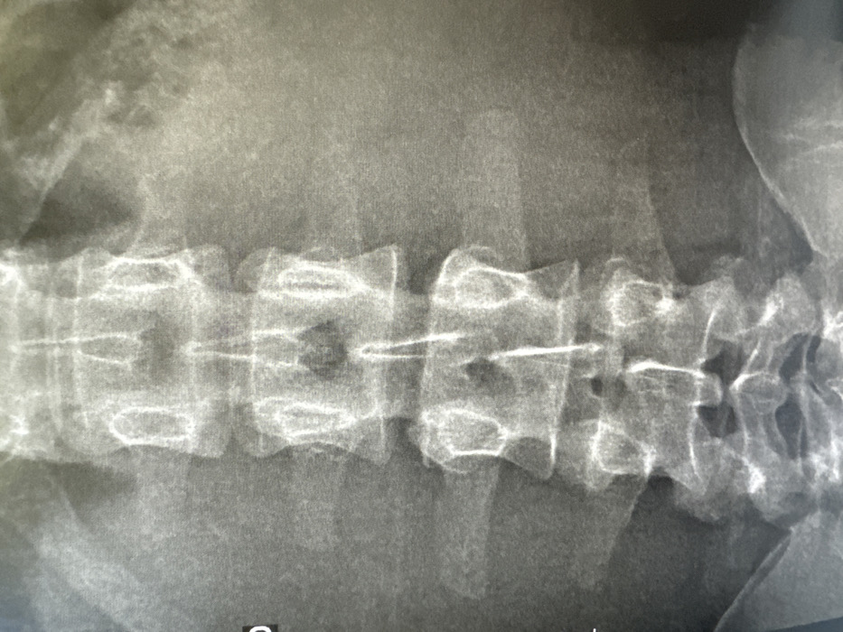
The image is an anterior-posterior (AP) radiographic view of a human lumbar spine, originating from the city of Vincennes, France. The X-ray displays five lumbar vertebrae, designated L1 through L5, along with the superior aspect of the sacrum and portions of the iliac bones forming part of the pelvis. The vertebral bodies appear generally intact, with discernible intervertebral disc spaces. A thin, linear, radiopaque object, consistent with a medical implant or catheter, is vertically oriented within the central canal region, extending from approximately the L1/L2 interspace down to the L4/L5 interspace. Minimal soft tissue shadows are visible on the periphery. The overall scene is a medical diagnostic image.
Loading AttnAds…
No transactions found