Sign in
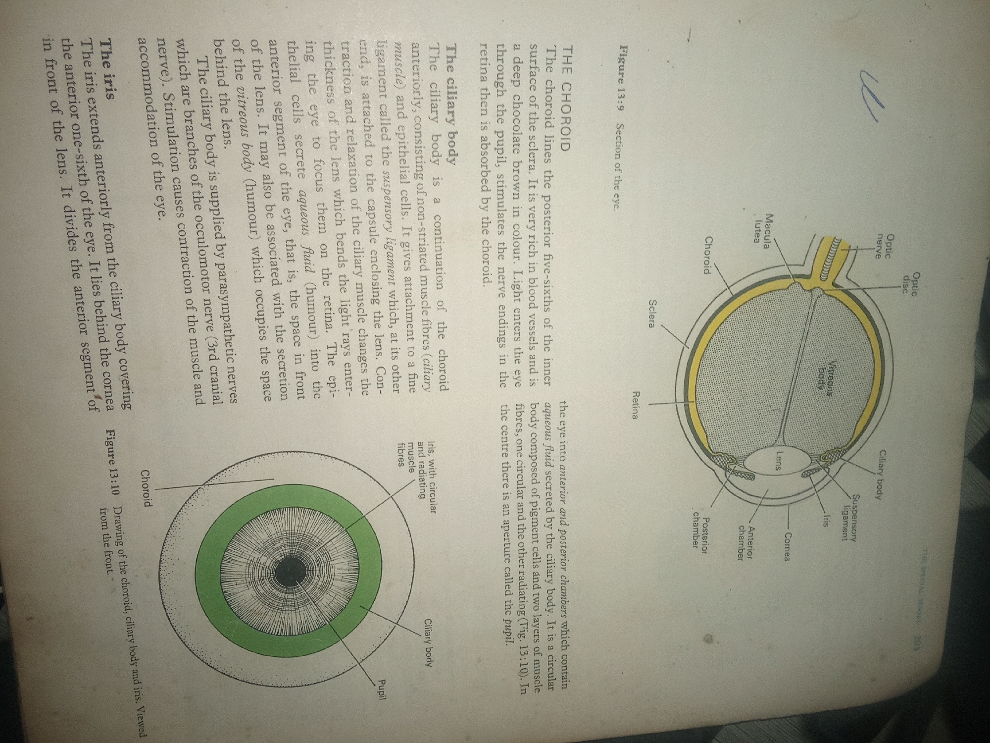
This media file is a photograph of an open textbook, displaying anatomical diagrams of the human eye and accompanying text. The page is likely from a biology or anatomy textbook, given the detailed illustrations and scientific terminology.

The main subjects are the anatomical structures of the eye, including the choroid, ciliary body, iris, lens, retina, optic nerve, optic disc, macula lutea, cornea, anterior chamber, posterior chamber, and vitreous body. The diagrams are rendered in color and black and white, with labels pointing to each part. One diagram is a cross-section of the eye, showing its internal layers, while the other is a frontal view of the iris, ciliary body, and choroid.

The visible text includes titles like "THE CHOROID," "The ciliary body," and "The iris," along with descriptive paragraphs explaining the functions and locations of these eye structures. There are also figure captions, such as "Figure 13:9 Section of the eye" and "Figure 13:10 Drawing of the choroid, ciliary body and iris. Viewed from the front." The text is in English.

There are no people or obvious signs of a specific time of day, weather, or emotion. The setting is clearly an academic or study environment, as indicated by the textbook format and scientific content. The location context provided (Jalingo, Nigeria) is not evident in the image itself, suggesting it is external information about where the photograph was taken or where the book is used.
FM-KpyEx1

Jun 30, 2026, 3:56 PM

Jalingo, Nigeria

Stake attention in this memory

curious
analytical
studious
objective
intellectual

This media file is a photograph of an open textbook, displaying anatomical diagrams of the human eye and accompanying text. The page is likely from a biology or anatomy textbook, given the detailed illustrations and scientific terminology. The main subjects are the anatomical structures of the eye, including the choroid, ciliary body, iris, lens, retina, optic nerve, optic disc, macula lutea, cornea, anterior chamber, posterior chamber, and vitreous body. The diagrams are rendered in color and black and white, with labels pointing to each part. One diagram is a cross-section of the eye, showing its internal layers, while the other is a frontal view of the iris, ciliary body, and choroid. The visible text includes titles like "THE CHOROID," "The ciliary body," and "The iris," along with descriptive paragraphs explaining the functions and locations of these eye structures. There are also figure captions, such as "Figure 13:9 Section of the eye" and "Figure 13:10 Drawing of the choroid, ciliary body and iris. Viewed from the front." The text is in English. There are no people or obvious signs of a specific time of day, weather, or emotion. The setting is clearly an academic or study environment, as indicated by the textbook format and scientific content. The location context provided (Jalingo, Nigeria) is not evident in the image itself, suggesting it is external information about where the photograph was taken or where the book is used.

Loading AttnAds…

transactions
revenues
stakers
Earliest
Latest
Highest stake

No transactions found

More from this user

More from Jalingo

FM-KpyEx1

Jun 30, 2026, 3:56 PM

Jalingo, Nigeria

Licensed under Creative Commons Attribution 4.0 International License. You are free to share and adapt this content with proper attribution.