Stake attention in this memory
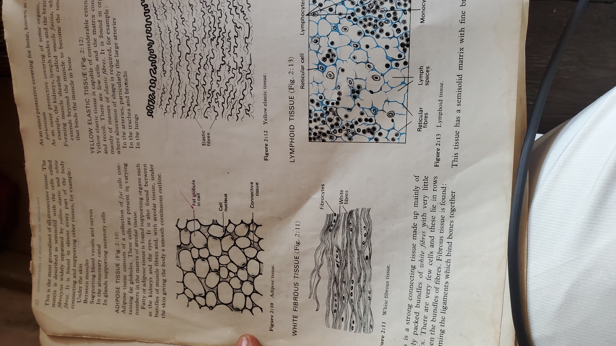
The image is a close-up of an open textbook page detailing different types of connective tissue. The page is divided into sections with accompanying diagrams. The top left section discusses "ADIPOSE TISSUE (Fig. 2:10)" with a diagram showing fat cells. A label points to "Fat globule in cell," "Cell nucleus," and "Connective tissue." To the right of the adipose tissue section, there's a description of "YELLOW ELASTIC TISSUE (Fig. 2:12)" accompanied by a diagram illustrating elastic fibers. Labels indicate "Elastic fibres." Below the adipose tissue diagram, the section on "WHITE FIBROUS TISSUE (Fig. 2:11)" is described, with a diagram showing parallel bundles of white fibers. Labels point to "Fibrocytes" and "White fibres." To the right of the white fibrous tissue section is "LYMPHOID TISSUE (Fig. 2:13)," with a diagram showing cells and spaces. Labels include "Reticular cell," "Lymphocytes," "Lymph spaces," and "Monocytes." The descriptions and diagrams are primarily in black and white, with some blue highlighting in the lymphoid tissue diagram. The text is printed, and there is a dark shadow line across the bottom of the image, suggesting the book is laid open. The page appears slightly yellowed, indicating age. The requested location in Jalingo, Nigeria, is not present in the image. The image is of a textbook and does not depict any real-world scenes or locations.
No transactions found